Without evidence of benefit, an intervention should not be presumed to be beneficial or safe.

- Rogue Medic

Another Sinus Tachycardia that F&B Medics Want to Shock


In Worst test question ever! – Maybe, I pointed out some of the problems with the way we “educate” people in EMS. The following question was provided with the rhythm above.
 

You are dispatched emergency traffic to the scene of a 24 yo F with “palpitations.” You arrive to find her pale, sweaty and lethargic. You palpate a radial pulse with an extreme rate. You hook her up to the monitor and find the following rhythm? You have a 45 minute transport time. Which of the following is the most appropriate initial treatment for this condition?

1.) Nitroglycerin 0.4mg SL
2.) Immediate synchronized cardioversion
3.) Adenosine 12mg Rapid IV push followed by 20cc NS bolus
4.) Epinephrine 1mg 1:10000 q-3-5m IVP


If a test question is so poorly written that there is no justifiable answer, why would anyone competent defend the question?

The answer is that we have dangerous people as “educators” and no really good method of eliminating them from EMS classrooms.

If there are no correct answers, can there be one best answer?

If there are no correct answers, can the most deadly answer possibly be the best answer?

We are supposed to be providing patient care, not making “educators” feel good about being dangerous.

Could the rhythm be atrial flutter?

It is not a bad idea to suspect flutter when the ECG rate is an even fraction of the ≈300 rate that is typical of flutter waves e.g. ≈75, ≈100, and ≈150)?

Is there another wave half way between the P waves seen in this rhythm, which would make both waves F waves?

I do not see any Flutter waves.


That is the same rhythm. Do you see any F waves?

If the rhythm is sinus, then cardioversion/defibrillation is not going to help, but can make things a lot worse.

We can produce anything from pain to death by shocking this rhythm.

Why would we do that to satisfy an incompetent “educator”?

This might be a good question to identify people who should not be trusted to care for patients. If we are willing to make a choice that is the equivalent of harming patients in order to come up with the one best answer right answer in a testing environment, what might we do when faced with a real patient, but a protocol that we think needs to be followed?

Is cardioversion the way to avoid the QA/QI/CYA spanking?

Should we be trying to figure out how to intervene?

Should we be trying to figure out when to intervene?

If the intervention is a heart-stopping dose of electricity (cardioversion), should we be looking for excuses to shock, or should we be trying to figure out if the patient is likely to benefit from that treatment?

What about this 80+ year old patient, who is pale, and lethargic, but is not confused?


Here is a different EMS 12 Lead Facebook post.
 

More ACLS…
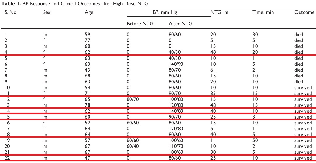
Your pt is a 54 yo male found in bed, with AMS. He looks pale, has BP of 84/52 with the rhythm below. No other hx is available right now. What’s the treatment?


The earlier post looked like a sinus tachycardia from a rhythm generator. With the HeartSim, to get a good fast narrow complex rhythm use atrial tachycardia and hit the faster button once or twice – that produces a rhythm that is not obviously sinus. There were 25 comments choosing from among the choices given, before there was a comment from someone smart enough to recognize that all of the choices were wrong.

Yes, the National Registry does encourage the fraud of one best answer. Why do medics choose a dangerous treatment, when we know it is dangerous?

We have been taught to choose try to figure out what would be most pleasing to the instructor/evaluator – not to do what is best for the patient.

Why aren’t the instructors/evaluators looking for what is best for the patient?

 
One of the reasons for fewer indefensible answers at EMS 12 Lead is that many of the people following EMS 12 Lead are already medics, nurses, and/or doctors, rather than students looking to please an instructor/evaluator.

This time, there were four good answers before anyone suggested cardioversion, but why are so many of us looking for excuses to use electricity to stop the hearts of patients?

.

Worst test question ever! – Maybe


Thank you to David Baumrind of EMS 12 Lead for linking to this here. It probably is not the worst test question ever, but it is very bad.

Read the question, figure out what your response would be, then scroll down for my explanation.
 

You are dispatched emergency traffic to the scene of a 24 yo F with “palpitations.” You arrive to find her pale, sweaty and lethargic. You palpate a radial pulse with an extreme rate. You hook her up to the monitor and find the following rhythm? You have a 45 minute transport time. Which of the following is the most appropriate initial treatment for this condition?

1.) Nitroglycerin 0.4mg SL
2.) Immediate synchronized cardioversion
3.) Adenosine 12mg Rapid IV push followed by 20cc NS bolus
4.) Epinephrine 1mg 1:10000 q-3-5m IVP

-Admin Paul

The original posting was from Exhausted Medic Students ‘R’ Us here.

Go read the original with its hundreds of comments.


*


*


*


*


*


*


*


*


*


*


*


All of the answers are completely wrong.
 

ST (Sinus Tachycardia) is the rhythm.

There are clear P waves with consistent PR intervals. It is faster than what some people expect to see from ST, but that is because many of us do not think about what we are learning in EMS.

It is true that the cardiology part of paramedic school is probably the toughest for most people, and we are overwhelmed with new information, but we should be very familiar with this rhythm.

Carry a patient up/down a flight of steps and you may have significant ST – maybe even faster than what is on this strip. If your heart rate is over 150, so what?

Before you have a chance to recover, use the pulse oximeter to measure your heart rate after carrying a patient. You are just checking the accuracy of the machine before applying it to the patient, or before reconnecting it to the patient.
 

1. Nitroglycerin is NOT indicated for palpitations.

NTG is not indicated even for a lot of palpitations. Do you have a protocol for NTG for palpitations?

Ask your medical director how much NTG should be given for palpitations, but don’t be surprised if you are expected to go through some scenarios to demonstrate that you would not really give NTG for palpitations.
 

2. Cardioversion is NOT indicated for sinus tachycardia.

Cardioversion is supposed to cause asystole. During that asystole, it is hoped that the sinus node will become the pacemaker for the patient’s rhythm.

SINUS tachycardia means that the sinus node is already the pacemaker.
 

Cardioversion of sinus tachycardia can only make things worse.
 

Cardioversion of sinus bradycardia can only make things worse.

Cardioversion of any sinus rhythm can only make things worse.
 

3. Adenosine is NOT indicated for sinus tachycardia.

The dose does not matter. The drug is not indicated.

No matter how wrong NTG is for palpitations, adenosine is worse.
 

4. Epinephrine is NOT indicated for sinus tachycardia with a pulse.

How much faster do we want this ST to be? Epinephrine can make it faster.
 

Maybe some people think that the choices should include a vagal maneuver.

No. That would also be wrong.

Calcium channel blocker?

Another wrong.

Beta blocker?

Wrong again.
 

No competent paramedic should attempt to justify any of these answers.

Maybe this is a question to find out just how incompetent people will be to satisfy an authority figure.

One horrible answer is –
 

As a paramedic instructor and a evaluator for National Registry…if my student didn’t cardiovert…I’m failing them.


Does the National Registry hire people this ignorant as evaluators?

Yes, but so does every other testing organization. Maybe this guy is lying about being an instructor and evaluator, but this is EMS and we like low standards.

A defender of cardioversion posted the ACLS tachycardia cheat sheet.
 

Click on image to make it larger.


Unfortunately, the cheat sheet does not state that we should not shock sinus tachycardia.

If all we know is the cheat sheet, we should consider a career change to explore the exciting world of fast food order fulfillment.

The text of the 2010 ACLS guidelines states –
 

ACLS professionals should be able to recognize and differentiate between sinus tachycardia, narrow-complex supraventricular tachycardia (SVT), and wide-complex tachycardia.[1]


A lot of people could not recognize an obvious sinus tachycardia.

Is that the fault of their instructors?

Yes and No.
 

Sinus tachycardia is among the rhythms listed that we are expected to be able to identify.
 

Synchronized cardioversion is recommended to treat (1) unstable SVT, (2) unstable atrial fibrillation, (3) unstable atrial flutter, and (4) unstable monomorphic (regular) VT. Shock can terminate these tachyarrhythmias by interrupting the underlying reentrant pathway that is responsible for them.[1]


Sinus tachycardia is not listed among the rhythms that should be shocked.

Here is the important part –
 

If judged to be sinus tachycardia, no specific drug treatment is required. Instead, therapy is directed toward identification and treatment of the underlying cause. When cardiac function is poor, cardiac output can be dependent on a rapid heart rate. In such compensatory tachycardias, stroke volume is limited, so “normalizing” the heart rate can be detrimental.[1]


We treat sinus tachycardia by treating the cause.

The cause of sinus tachycardia is never lack of cardioversion.
 

A good test near the end of the cardiology section of paramedic school might include this question to find out if the students have learned anything.

All of the choices are wrong.
 

In medicine, there is not one best answer for all patients.
 

Anyone who says differently is selling something.

Footnotes:

[1] Tachycardia
2010 American Heart Association Guidelines for Cardiopulmonary Resuscitation and Emergency Cardiovascular Care Science
Part 8: Adult Advanced Cardiovascular Life Support
Part 8.3: Management of Symptomatic Bradycardia and Tachycardia
Cardioversion and Regular Narrow-Complex Tachycardia

.

Woman with Risks for Torsades de Pointes Dying within Hours of Leaving the Emergency Department


I don’t expect to see this as a headline anywhere, but this possible cause of death something we should be aware of.

Abdominal pain in a patient with many comorbidities. She is given medication and later is found dead at her home.

What drugs was she taking?
 

a Potentially proarrhythmic drugs as classified by the Arizona Center for Education and Research on Therapeutics (www.qtdrugs.org).
b Given during emergency department visit.[1]


What risk factors did she have?
 

a Risk factors present in case study.[1]


The 12 lead obtained in the ED (Emergency Department) shows a bradycardia with a heart rate of 58 beats per minutes. Bradycardia probably should have been included in the risk factors in this case.

What treatment did she receive that increased her risk of TdP (torsades de pointes)?


Ondansetron (Zofran)
 

On September 15, 2011, the FDA issued a Medwatch Safety Alert for Zofran (ondansetron) in patients with congenital Long QT syndrome, a heart arrhythmia. The FDA further required GlaxoSmithKline to conduct a thorough QT study to determine the degree to which Zofran may cause QT interval prolongation.[1] On June 29, 2012, the FDA issued an FDA Drug Safety Communication Update entitled New information regarding QT prolongation with ondansetron (Zofran).

The 32-mg high dose of ondansetron (Zofran) has been pulled from the market by the FDA because of concerns about cardiac problems.[15][2]


The high dose of 32 mg is more than would usually be given by EMS or in the ED.

In this case, the ondansetron was 8 mg given orally in the ED, so this was a much smaller dose.

What is QT segment prolongation?


Image credit.

There is a problem with the image. The ventricles contract during the QRS complex, not during the T wave.
 

Let’s see some torsades.


Click on images to make them larger.[3]
 

How do we know that it is TdP?

Because of the long QT segment in the beats preceding the VT (Ventricular Tachycardia).

Does all torsades go away on its own, as the above example did?

No.


A medical screening examination was conducted and 8 mg of orally disintegrating ondansetron (Zofran) was administered for persistent nausea and vomiting. A 12-lead electrocardiogram (ECG) completed at triage (Figure 1) was remarkable for left ventricular hypertrophy and QT interval prolongation.[1]


This is a patient who should be on a monitor, not necessarily because of the proarrhythmic effects of the drugs she is already taking, but because of the combination with the proarrhythmic drug she has been given in the ED.


Shortly thereafter, the patient self-discharged from the emergency department before receiving definitive treatment. Upon making a follow-up phone call, it was discovered that the patient had been found unresponsive in bed approximately 4 hours after leaving the emergency depart[1]


Was it the Zofran?

Maybe, but if it was, the ondansetron may only be the straw that broke the camel’s back.

Does that mean that the risks should have been ignored?

No.

Many of the patients we see are the most fragile people in society and we are seeing them when they are at their greatest vulnerability to adverse treatment effects.

The FDA has warned about the QT prolonging effects of ondansetron, so we cannot claim that we could not have known. I have written about this before.[4],[5]

We should be looking for reasons why we should not be giving treatments.

EMS operates under protocols that may state –

If condition X is present, give treatment A, then give treatment B.

ED treatment can be just as protocol driven as EMS treatment.

We have drugs that can be dangerous under certain circumstances.

Should we give any drug without considering the possible drug interactions and adverse events?

If we are not aware of the drug’s possible drug interactions and adverse events, should we be permitted to give the drug?

Ondansetron is one of the drugs I give frequently, but I need to remind myself to consider the possible QT prolonging effects and to look for other QT prolonging drugs and medical conditions.

What other drugs do I carry that can cause QT prolongation?

Amiodarone (Nexterone, Cordarone) is the only drug I carry that is on the Drugs with a Risk of Torsades de Pointes list. Even ondansetron is not on this list. There is less evidence that ondansetron causes torsades, than there is that amiodarone causes torsades.
 

Substantial evidence supports the conclusion that these drugs prolong the QT interval and have a risk of TdP when used as directed in labeling.[6]


Oxytocin (Pitocin) and ondansetron are on the Drugs with a Possible Risk of Torsades de Pointes list.
 

Substantial evidence supports the conclusion that these drugs cause QT prolongation but there is insufficient evidence that they, when used as directed in labeling, have a risk of causing TdP.[6]


Diphenhydramine (Benadryl) is the only drug I carry that is on the Drugs with a Conditional Risk of Torsades de Pointes list.
 

Substantial evidence supports the conclusion that these drugs prolong the QT interval and have a risk of TdP but only under certain known conditions (e.g. excessive dose, drug interaction, etc.).[6]


All of these drugs are generally considered to be safe, because we are ignorant of the adverse events they can cause. TdP is only one adverse event. Amiodarone has other proarrhythmic effects, can cause hypotension,

Then there are the many drugs that may interact with these drugs to prolong the QT segment.

Antibiotics, psychiatric medications (for all kinds of psychiatric conditions – psychosis to depression), erectile dysfunction drugs,

Then why aren’t we seeing large numbers of dead bodies?

These patients have other medical conditions that may lead to death without any TdP or there may not be much TdP caused by these drugs. We do not know.

We do know that thousands, even tens of thousands of patients can die without anyone noticing that the deaths are the effect of a drug.[7]

For example –
 

She was pronounced dead at the scene by emergency care providers. Because of her extensive medical history, the woman’s family declined an autopsy, and her primary physician attributed the death to complications of diabetes mellitus, end-stage renal disease, and hypertension.[1]


What was the cause of death?

We do not know.

Given the number of risk factors for TdP, is TdP a likely cause?

Torsades de pointes is no less likely a cause of death than anything listed by the her primary care physician on the death certificate.

This isn’t a multiple choice exam, where someone thinks that there is some mythological one best answer.

This is the real world and all of these conditions probably significantly contributed to her death.

Was ondansetron the final straw?

Maybe.

Footnotes:

[1] Woman with Risks for Torsades de Pointes Dying within Hours of Leaving the Emergency Department.
Pickham D, Sickler K.
J Emerg Nurs. 2011 Dec 2. [Epub ahead of print] No abstract available.
PMID: 22137882 [PubMed – as supplied by publisher]

[2] Ondansetron
Wikipedia
Adverse effects
Article

[3] Etiology, warning signs and therapy of torsade de pointes. A study of 10 patients.
Keren A, Tzivoni D, Gavish D, Levi J, Gottlieb S, Benhorin J, Stern S.
Circulation. 1981 Dec;64(6):1167-74.
PMID: 7296791 [PubMed – indexed for MEDLINE]

Abstract with link to Free Full Text Download in PDF format from Circulation

[4] Ondansetron (Zofran) Warning for QT Prolongation – is Amiodarone next? – Part I
Mon, 02 Jul 2012
Rogue Medic
Article

[5] Ondansetron (Zofran) Warning for QT Prolongation – is Amiodarone next? – Part II
Thu, 05 Jul 2012
Rogue Medic
Article

[6] TdP drug lists
AZCERT.org is now CredibleMedsTM
Web page with links to all lists as pop ups.

[7] Mortality and morbidity in patients receiving encainide, flecainide, or placebo. The Cardiac Arrhythmia Suppression Trial.
Echt DS, Liebson PR, Mitchell LB, Peters RW, Obias-Manno D, Barker AH, Arensberg D, Baker A, Friedman L, Greene HL, et al.
N Engl J Med. 1991 Mar 21;324(12):781-8.
PMID: 1900101 [PubMed – indexed for MEDLINE]

Free Full Text Article from N Engl J Med.

.

How do we measure the QT segment when there are prominent U waves?

ResearchBlogging.org
 

This ECG has large T waves, U waves, and P waves, but where does one end and the other begin?

When measuring the QT segment, where do we measure the end of the QT segment and why?

Click on the image to make it larger.
 

Fig. 1 Electrocardiogram demonstrating the phenomenon of T-U fusion.[1]


Leads V2 – V4 are the most distinct, but the T wave and the U wave are not distinct from each other. They are connected with no isoelectric line between them. Therefore, the QT segment becomes more of a QTU segment. If the T wave and the U wave did not merge, or fuse, then the QT segment would not include the U wave.
 

Amiodarone is a widely used antiarrythmic drug for various atrial and ventricular arrhythmias. It has the potential to cause prolongation of the QT interval, which, in turn, can increases the incidence of torsade de pointes. Amiodarone is also one of the causes of prominent U waves. The presented case exemplifies the phenomenon of amiodarone-induced T-U fusion and QT prolongation.[1]


We seem to forget that any antiarrhythmic drug is also capable of causing arrhythmias. If we alter the conduction system, we can make things better, we can make things worse, or we can produce a combination of both. It is possible to have no effect, but this is more likely just our lack of awareness of the effects we are producing.
 

Amiodarone is also one of the causes of a prominent U wave in the surface electrocardiogram [2]. It blocks the delayed rectifier potassium current, thereby delaying phase 3 of action potential. This repolarization delay may distort T waves and/or produce prominent U waves[3,4].[1]


Amiodarone and torsades is a significant, but not well known problem.
 

Proarrhythmia
Like all antiarrhythmic agents, amiodarone I.V. may cause a worsening of existing arrhythmias or precipitate a new arrhythmia. Proarrhythmia, primarily torsades de pointes (TdP), has been associated with prolongation by amiodarone I.V. of the QTc interval to 500 ms or greater. Although QTc prolongation occurred frequently in patients receiving amiodarone I.V., torsades de pointes or new-onset VF occurred infrequently (less than 2%). Patients should be monitored for QTc prolongation during infusion with amiodarone I.V. Combination of amiodarone with other antiarrhythmic therapy that prolongs the QTc should be reserved for patients with life-threatening ventricular arrhythmias who are incompletely responsive to a single agent.
[2]


Here is an explanation of how to measure the QT segment when there is fusion with a prominent U wave.


Fig. 2 Panel A represents a magnified segment of lead V 2 of the presented electrocardiogram which demonstrates T, U, and P waves and the phenomenon of T-U fusion.[1]


Fig. 2 Panel B illustrates the application of the maximum slope intercept method to the U wave to calculate the QTc; the QTc (QT/√RR) is prolonged to 510 milliseconds.[1]


If the T wave reaches the isoelectric line, we measure the end of the QT segment at that point. If the T wave runs into the U wave, we should draw the steepest (closest to vertical) line that we can along the descending side of the U wave (Figure 2B).

This makes for a significantly longer QT segment. The longer the QT segment, the greater the risk for torsades appears to be. Torsades is not our friend.[3],[4]

Footnotes:

[1] Amiodarone-induced T-U fusion.
Omar HR.
Am J Emerg Med. 2012 Nov;30(9):2081.e1-2. doi: 10.1016/j.ajem.2011.10.024. Epub 2011 Dec 26. No abstract available.
PMID: 22205017 [PubMed – in process]

[2] AMIODARONE HYDROCHLORIDE injection, solution
[Bedford Laboratories]

DailyMed
Warnings
FDA Label

[3] 47 Year Old Male CC: Crushing Chest Pain
August 11, 2011
EMS 12 Lead
Article

[4] 47 Year Old Male CC: Crushing Chest Pain – Conclusion
August 17, 2011
EMS 12 Lead
Article

Omar, H. (2012). Amiodarone-induced T-U fusion The American Journal of Emergency Medicine, 30 (9), 20810-208100 DOI: 10.1016/j.ajem.2011.10.024

.

Should the AHA reintroduce the ‘Indeterminate’ class of ACLS recommendations?


In 2005 the AHA (American Heart Association) used more categories for their drug recommendations. The recent very poor showing of epinephrine in resuscitation studies, and the lack of evidence of benefit in earlier studies, suggest that Class IIb is not an appropriate classification of epinephrine.

Should the AHA reintroduce the Indeterminate class that was in the 2005 guidelines?

Can we justify continuing the Class IIb recommendation?

Where are the people who survived because of epinephrine?

With hundreds of thousands of cardiac arrest patients treated with epinephrine each year, why can’t we find any evidence of improved survival with epinephrine?

All we need is –

Benefit Risk

Where is the benefit?

Why are we satisfied with such pathetic results?
 

Table 3.
Applying Classification of Recommendations and Level of Evidence

Class I
Benefit >>> Risk
Procedure/treatment or diagnostic test/assessment should be performed/administered.

Class IIa
Benefit >> Risk
It is reasonable to perform procedure/administer treatment or perform diagnostic test/ assessment.

Class IIb
Benefit Risk
Procedure/treatment or diagnostic test/assessment may be considered.

Class III
Risk Benefit
Procedure/treatment or diagnostic test/assessment should not be performed/administered. It is not helpful and may be harmful.

Class Indeterminate.
• Research just getting started
• Continuing area of research
• No recommendations until further research (eg, cannot recommend for or against)[1]


Is epinephrine research just getting started?

After over half a century of use, there is only a single prospective randomized, placebo-controlled study of the effects of epinephrine on outcomes. That is from last year and the study was decimated by politicians and the press claiming that it would be unethical to deprive patients of this untested treatment.
 

Is epinephrine a continuing area of research?

I don’t know, but it should be. We should not continue to use epinephrine without continuing research.
 

Is epinephrine deserving of any recommendation without further research?

So far, there is only evidence of improvement in surrogate endpoints. surrogate endpoints are things that we measure because a study is too small to show improved survival.
 

Here is the evidence of of what happens to survival with epinephrine.


I added the two most recent studies to this, since they were not yet published when the review of vasopressors was published.[2] The quality and outcome of the added studies is my interpretation, but I think that others will rank them similarly.

The Hagihara study is more than 100 times larger than the combined numbers of all of the studies that came before it.

The Hagihara study is not positive.

Why aren’t we demanding evidence that we are not harming patients?
 

We don’t know enough to know how dangerous epinephrine is.
 

That is the best that can be stated about epinephrine in cardiac arrest.

Isn’t it time for us to figure out what we are doing?

We should have found out whether epinephrine works before we started using it on everyone dead, but we don’t have to continue that mistake.

Our patients need to know. Does epinephrine improve survival from cardiac arrest?

Footnotes:

[1] Table 3. Applying Classification of Recommendations and Level of Evidence
2005 American Heart Association Guidelines for Cardiopulmonary Resuscitation and Emergency Cardiovascular Care
Part 1: Introduction
Table 3

I have modified this table solely for the purpose of clarity of presentation, by modifying color and font. None of the words have been changed.

[2] Vasopressors in cardiac arrest: a systematic review.
Larabee TM, Liu KY, Campbell JA, Little CM.
Resuscitation. 2012 Aug;83(8):932-9. Epub 2012 Mar 15.
PMID: 22425731 [PubMed – in process]

CONCLUSION: There are few studies that compare vasopressors to placebo in resuscitation from cardiac arrest. Epinephrine is associated with improvement in short term survival outcomes as compared to placebo, but no long-term survival benefit has been demonstrated. Vasopressin is equivalent for use as an initial vasopressor when compared to epinephrine during resuscitation from cardiac arrest. There is a short-term, but no long-term, survival benefit when using high dose vs. standard dose epinephrine during resuscitation from cardiac arrest. There are no alternative vasopressors that provide a long-term survival benefit when compared to epinephrine. There is limited data on the use of vasopressors in the pediatric population.

.

Is Digoxin a Killer?

ResearchBlogging.org
 

From the London Science Museum – Bottle of digoxin tablets, ‘Tabloid’ brand, London, England, 1884-1924.

Digoxin (Lanoxin) is an antiarrhythmic drug, which means that it is also a proarrhythmic drug. Any drug that affects the heart’s conduction system can produced changes that are bad, good, or a combination of the two. Digoxin has been associated with a higher death rate, but is that because it is prescribed to sicker patients?
 

Digoxin has been used worldwide for decades to achieve rate control in patients with AF. Its use in heart failure (HF) dates back centuries but remains controversial,5 due to its narrow therapeutic index and a potential to contribute to life-threatening ventricular tachyarrhythmias and severe bradyarrhythmias.6,7 [1]


Digoxin is an antiarrhythmic, but a Class V antiarrhythmic. It doesn’t fit in anywhere, so it is in the none of the above class. Digoxin also is a rare inotrope (causes more forceful contraction of the heart), while generally not raising the heart rate. If the heart is already in failure, increasing heart rate to increase cardiac output may not be a good idea, which makes digoxin appealing and possibly beneficial in HF.
 

Digoxin is known to slow heart rates and potentiate bradyarrhythmias25 through its parasympathetic effect on the AV node, but has little effect on fast ventricular rates in the setting of enhanced sympathetic tone.26 Therefore, digoxin is not the ideal choice to control rapid ventricular rates in most patients.[1]


AFFIRM was looking at whether rate control or rhythm control produced a better outcome for patients, not whether digoxin is beneficial, or harmful. The data are relevant for evaluating the possible risks from digoxin.


The AFFIRM trial design, baseline characteristics, and results have been published previously.4 In brief, the study enrolled 4060 patients with AF considered at high risk for stroke. These patients were randomized to rate control vs. rhythm control over a 4-year period with a mean follow-up of 3.5 years.[1]


This study looks at the data from AFFIRM and EHRs (Estimated Hazard Ratios) for many risk factors with and without digoxin.
 

Digoxin was associated with increased all-cause (EHR 1.41, 95% CI 1.19–1.67, P < 0.001) and cardiovascular mortality (EHR 1.35, 95% CI 1.06–1.71, P = 0.016) after controlling for clinical and demographic variables, as well as propensity scores. Similarly, digoxin was associated with an increase in arrhythmic deaths (EHR 1.61, 95% CI 1.12–2.30, P = 0.009);[1]


This graph shows the progressive divergence of the mortality rates which should be expected if digoxin increases the death rate.
 

Click on images to make them larger.
 

The one thing I did not like about this paper is the way they organized the data. If you look at the original tables, they are not organized in a way that helps to understand the relationships among different risk factors by organizing them according to their association with mortality. I rearranged the tables by p values to try to make those associations easier to see.


These all met statistical significance of a p value of less than 0.05, arranged from the weakest association to the strongest association within 6 months of adding digoxin. For example, patients with a history of cardiomyopathy did not do well on digoxin.


These are from the same the risk factors, but these are the ones that did not meet statistical significance. Prior interventional procedure actually trends toward a benefit with digoxin, while a Permanent pacemaker seems to be the least likely to be affected by digoxin.


These are Cardiovascular mortality and All-cause mortality, based on whether they were taking digoxin on their last recorded visit, rather than just within 6 months of entering the study.

I should have arranged these by All-cause mortality with digoxin, but some people would claim that digoxin only has cardiovascular effects. The first table is of the variables that are not statistically significant.


These are the variables that reached statistical significance with digoxin. A p value of <0.05 is a result considered to have a less than 5% chance of being due to chance. If the original study were redone 20 times, there is the possibility that the results would come out this way purely due to chance one time. A p value of <0.0001 is a result considered to have a less than 0.01% chance of being due to chance. If the original study were redone 10,000 times, there is the possibility that the results would come out this way purely due to chance one time. Not as far fetched as the Powerball odds, but the lottery far fewer variables. Each has roughly a 0.000000005714 chance of winning.[2]
 

Comparing the EHR with all covariates present to that with none present, 62.6% of digoxin over-mortality cannot be attributed to confounding from the covariates.[1]


That is nice and simple.

There are plenty of excuses, such as being prescribed to sicker patients, but they do not appear to have any validity.
 

Patients without CHF or low EF lack the neurohormonal and inotropic derangements that may improve with digoxin, while remaining exposed to its potential deleterious effects such as proarrhythmia and bradycardia. In the AFFIRM trial, digoxin was utilized to meet the stringent rate control strategy requirement (resting heart rate <80 b.p.m. and exercise heart rate <110 b.p.m.), usually in combination with other atrioventricular (AV) nodal blockers such as beta-blockers or calcium-channel blockers. Indeed, digoxin was used as monotherapy for rate control in only 17% of patients.21 In those patients, higher doses of digoxin with an increased risk for toxicity may have been used to achieve the stringent rate control goal, as high serum levels of digoxin were encouraged in the AFFIRM protocol (>1.0 ng/mL).[1]


They were not able to adjust for the large doses of digoxin used in AFFIRM, so that may be a significant contributor to the elevated death rate.


The largest trial to examine the safety of digoxin in patients with HF, the DIG study, excluded patients with AF.10 In that trial, patients were randomized to digoxin vs. placebo. Digoxin was found to have a neutral effect on the all-cause mortality (EHR 0.99; 95% CI 0.91–1.07; P = 0.80). However, it is important to note that real-world patients, including those in AFFIRM, are not routinely subject to the close follow-up and frequent monitoring of serum digoxin concentrations mandated in the DIG study. It is possible that such strict monitoring is required to ensure safety. Further analysis of the DIG trial data demonstrated that digoxin’s beneficial effect applied only to patients in SR with low serum digoxin drug levels (<0.9 ng/mL).7,32 [1]


If the benefits of digoxin are limited to patients with a SR (Sinus Rhythm) and only at low doses of digoxin, maybe digoxin should have very limited indications.
 

While digoxin’s positive neurohormonal effects in HF patients may be attenuated or lost when beta-blockers are concomitantly prescribed, the association between digoxin and mortality observed in our study was independent of beta-blocker use.[1]


Even beta-blockers do not seem to protect against digoxin.

Is the best way to avoid killing patients to use less digoxin and to only use digoxin for very specific indications?
 

Go read the full paper. I don’t know how long it will be free as Advanced Access.

Footnotes:

[1] Increased mortality among patients taking digoxin-analysis from the AFFIRM study.
Whitbeck MG, Charnigo RJ, Khairy P, Ziada K, Bailey AL, Zegarra MM, Shah J, Morales G, Macaulay T, Sorrell VL, Campbell CL, Gurley J, Anaya P, Nasr H, Bai R, Di Biase L, Booth DC, Jondeau G, Natale A, Roy D, Smyth S, Moliterno DJ, Elayi CS.
Eur Heart J. 2012 Nov 27. [Epub ahead of print]
PMID: 23186806 [PubMed – as supplied by publisher]

Free Full Text Advanced Access from European Heart Journal

[2] Why playing Powerball once is enough – Commentary: Loading up on tickets won’t up your odds — or your fun
November 28, 2012
Chuck Jaffe
MarketWatch
Article

The difference in the odds of winning if you buy a ticket, or a hundred tickets, is essentially indistinguishable from the odds if you don’t buy any ticket at all.

Whitbeck, M., Charnigo, R., Khairy, P., Ziada, K., Bailey, A., Zegarra, M., Shah, J., Morales, G., Macaulay, T., Sorrell, V., Campbell, C., Gurley, J., Anaya, P., Nasr, H., Bai, R., Di Biase, L., Booth, D., Jondeau, G., Natale, A., Roy, D., Smyth, S., Moliterno, D., & Elayi, C. (2012). Increased mortality among patients taking digoxin-analysis from the AFFIRM study European Heart Journal DOI: 10.1093/eurheartj/ehs348

.

Is a half a bottle of nitro too much for a single dose?

 
In what I last wrote about the emergency treatment of CHF (Congestive Heart Failure) and furosemide (Lasix – frusemide in Commonwealth countries) being a bad drug,[1] I also mentioned what Peter Canning had written about Hero Medic and a very high dose of NTG (NiTroGlycerin – GTN GlycerylTriNitrate in Commonwealth countries).

How much NTG?
 

The hero medic opened the nitro, pulled open the patient’s mouth and poured in what seemed like half the container. The hero medic closed the patient’s mouth, and then told the new medic. “You should be all set.”[2]


What seemed like half the container of something we are warned not to give chest pain patients more than one at a time and never more than 3 tabs of – ever!

That does seem like a lot, but how much is half of a bottle?


How much is a lot?


For the typical bottle pictured, the label states that the total is 25 of the 0.4 mg tabs.

That is a total of 10 mg. Half of that is easy math – 5 mg.

What seemed like half the container is probably less than half, but it was just such a shocking visual for the narrator, that all he could do was estimate.

Is 5 mg a large dose?

Yes.

Is 5 mg a dangerous dose?

That depends on the patient presentation.
 

a patient in severe pulmonary edema who was circling the drain[2]


I would like to know his blood pressure, but even hypotensive CHF patients do well with huge doses of NTG.

The patients below were more than just circling the drain.

All were hypotensive, or pulseless.


Click on the image to make it larger.[3]


Almost all of the massive dose NTG patients survived.

The highest blood pressure before NTG was 80/70.

The lowest dose of NTG was 1 mg – and that was the only patient who received less than 5 mg NTG.

Almost every hypotensive patient received much more than 5 mg NTG.

Is 5 mg NTG a dangerous dose?

Probably not.

Is 5 mg a scary dose?

Of course. We have been trained to be scared of NTG.

That dose is just much larger than most people are comfortable with, because of fear and inexperience.
 

You can talk about the hero medic acting off protocol and being a cowboy, etc, but the story still resonates for me.[2]


Would it be appropriate to just drop in, give a large dose of anything, and then not stick around?

No. If the original medic is not comfortable with that treatment, the Hero Medic should be there to hold the nitrophobic medic’s hand and reassure the medic to follow his assessment, rather than to follow his anxiety. That is the way we learn.

The story of Hero Medic is told through hyperbole – exaggeration to make an important point.

I have done this with others. Look, not only am I giving a much larger dose than you claim is safe – I am going to add a few more tabs – just to make a point.

If the scare stories about NTG were true, the patients would bottom out their blood pressures, but it is only the witnesses who bottom out their blood pressures, due to over-stimulation of their vagal nerves. The patients improve.

Some people learn from this clear demonstration that they have been lied to, but others choose to believe the dogma even after seeing clear evidence that the dogma is a lie.

Doctors, nurses, medics, et cetera. They are equally susceptible to dogma.

Is the Hero Medic a cowboy?

No, the Hero Medic s a teacher.

We need to be smart enough to learn.

Footnotes:

[1] Lasix Kills: Better Therapy for CHF
Wed, 07 Nov
Rogue Medic
Article

[2] NTG and the Hero Medic
Street Watch
November 7, 2012
Article

[3] High dose nitroglycerin treatment in a patient with cardiac arrest: a case report.
Guglin M, Postler G.
J Med Case Reports. 2009 Aug 10;3:8782.
PMID: 19830240 [PubMed]

Free Full Text from PubMed Central

.

What is the basis for post-resuscitation treatment recommendations?

We spend a lot of time worrying about how to get a pulse back. We spend so much time on getting that pulse back, that some of us think that producing a pulse is what matters.

Once we have a pulse back, whether using only evidence-based treatments (continuous compressions and defibrillation) or after throwing in some of the witchcraft that does not belong in the guidelines (ventilations, intubation, epinephrine, vasopressin, norepinephrine, phenylephrine, amiodarone, lidocaine, magnesium), we have no good evidence to guide treatment.
 

There is no proven benefit or harm associated with administration of routine IV fluids or vasoactive drugs (pressor and inotropic agents) to patients experiencing myocardial dysfunction after ROSC.[1]


This clearly rules out having any treatments categorized as Class I or even as Class IIa, but the AHA (American Heart Association) does not follow its rules for evaluation evidence. In the paragraph following the quote above, the guidelines state –
 

Fluid administration as well as vasoactive (eg, norepinephrine), inotropic (eg, dobutamine), and inodilator (eg, milrinone) agents should be titrated as needed to optimize blood pressure, cardiac output, and systemic perfusion (Class I, LOE B).[1]


Where is the evidence?
 

There is a paucity of data about which vasoactive drug to select first, although providers should become familiar with the differing adverse effects associated with these drugs, which might make a particular agent more or less appropriate for a specific patient.153 [1]


In other words, we should not select drugs because they are good, but because they do not have a lot of evidence of harm, or because we are comfortable with the excuses for the harm these drugs cause.

When presented with this kind of advice, we should always ask, What evidence do we have that any of these harmful treatments provide more benefit than harm?

In EMS, dopamine seems to be the drug of choice for shock (inadequate tissue perfusion, usually with hypotension).
 

Although low-dose dopamine infusion has frequently been recommended to maintain renal blood flow or improve renal function, more recent data have failed to show a beneficial effect from such therapy161,162 [2]

Are we asking the right questions?

Is vasoconstriction the solution to shock?

Is vasodilation the problem?
 

High doses of NTG were used in 22 patients, including 14 patients with acute MI and eight patients with advanced HF. All patients had critically low BP measured by cuff, and 18 had an unmeasurable BP and pulse.[3]


The doses shown are considered to be huge – even for patients who have very high blood pressures.

These patients do not have any blood pressure, or they have such very low blood pressure that it cannot be measured. The treatment is NTG (NiTroGlycerin or GTN – GlycerylTriNitrate in Commonwealth countries).

Almost any ACLS (Advanced Cardiac Life Support) instructor will tell you exactly what the result will be – death and course failure for refusal to accept the dogma of hypotension plus NTG = death.

I underlined all of the patients who received 25 mg NTG or more. A standard NTG tab is 0.4 mg. All of the underlined patients received an amount of IV (IntraVenous) NTG that is more than 60 times higher than the standard NTG dose.

In all of these patients, the blood pressure was too low to be obtained.


Click on the image to make it larger.


Almost all of the massive dose NTG patients survived.

If we are to believe the hypothesis that vasopressors are necessary to treat shock, improvement after NTG does not make sense.

If we are to believe the hypothesis that vasopressors are necessary to treat shock, improvement after huge NTG does is not possible.

Maybe we need to reconsider our beliefs.

We have developed a bias that is unduly influencing our treatment of our least stable patients.

We have developed a bias that is unduly influencing our treatment of our most easily harmed patients.

Footnotes:

[1] Vasopressors
2010 American Heart Association Guidelines for Cardiopulmonary Resuscitation and Emergency Cardiovascular Care
Part 9: Post–Cardiac Arrest Care
Vasoactive Drugs for Use in Post–Cardiac Arrest Patients
Free Full Text from Circulation

[2] Table 2. Common Vasoactive Drugs
2010 American Heart Association Guidelines for Cardiopulmonary Resuscitation and Emergency Cardiovascular Care
Part 9: Post–Cardiac Arrest Care
Vasoactive Drugs for Use in Post–Cardiac Arrest Patients
Free Full Text from Circulation

[3] High dose nitroglycerin treatment in a patient with cardiac arrest: a case report.
Guglin M, Postler G.
J Med Case Reports. 2009 Aug 10;3:8782.
PMID: 19830240 [PubMed – in process]

Free Full Text from PubMed Central . . . . . Free Full Text PDF from PubMed Central

.