Without evidence of benefit, an intervention should not be presumed to be beneficial or safe.

- Rogue Medic

Do Emergency Physicians Intubate Often Enough to Maintain Competency?


    There is a study of the frequency of intubation among emergency physicians in the current Annals of Emergency Medicine. This study is accompanied by a discussion, which unfortunately does not question the assumption that intubation improves outcome. There is very little evidence to suggest that intubation improves outcomes. That evidence is only using paramedics with the highest success rates – much higher than your average paramedic.

 
Greater intubation experience in paramedics is associated with improved patient outcomes2; does a similar relationship exist for emergency physicians?[1]


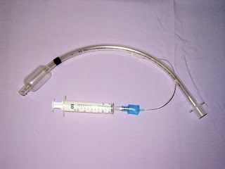
Image credit.

The unquestioned assumption is that excellent intubation performance improves outcomes, rather than that excellent intubation performance causes less harm than average intubation performance, or below average performance. We do not have any good evidence to support the wishful thinking that paramedics, or even much more experienced emergency physicians, improve outcomes by intubating patients. We just assume this, because we don’t really want to know. If we decide to be honest and actually find out the effect of intubation, how will we handle it if the results show that we are harming more patients than we are helping?

The Cardiac Arrhythmia Suppression Trial was only started because the proponents of the different antiarrhythmics (encainide, flecainide, and moricizine) wanted to prove that their drug was better than all of the rest. They even agreed to include a placebo arm, although the doctors did not like the idea of depriving patients of such beneficial treatment.

 
CONCLUSIONS: There was an excess of deaths due to arrhythmia and deaths due to shock after acute recurrent myocardial infarction in patients treated with encainide or flecainide.[2]
 

People who had frequent ectopic heart beats – PVCs (Premature Ventricular Contractions) after a heart attack were more likely to die than people who did not have frequent PVCs. The obvious solution – the equivalent of intubation and blood-letting – was to give drugs that will get rid of the PVCs. The problem is that the PVCs were not the problem. The PVCs were just a sign of the problem. The drugs made the actual problem with the heart worse, while making the heart appear to be better. The same is true of blood-letting and may be true of intubation. Abundant evidence for the obvious benefits of blood-letting are quoted in the footnotes.[3]

If intubation is harmful, do we want to know?

If intubation by the average paramedic is harmful, do we want to know?

If intubation by the average emergency physician is harmful, do we want to know?

It isn’t as if we take intubation seriously. If we did take intubation seriously, we would practice much, much more than we do. In stead, we make excuses for failing to practice something that we claim is life-saving, because we are too arrogant to admit that practice is important to develop and maintain any skill.

Practicing on even the most basic mannequin should be done before every shift, whether you are a paramedic or an emergency physician. Unless you have a 99%, or better, success rate on hundreds of patients.

Footnotes:

[1] Intubation by Emergency Physicians: How Often Is Enough?
Kerrey BT, Wang H.
Ann Emerg Med. 2019 Dec;74(6):795-796. doi: 10.1016/j.annemergmed.2019.06.022. Epub 2019 Aug 19. No abstract available.
PMID: 31439364

The article above is commentary on the article below:

Procedural Experience With Intubation: Results From a National Emergency Medicine Group.
Carlson JN, Zocchi M, Marsh K, McCoy C, Pines JM, Christensen A, Kornas R, Venkat A.
Ann Emerg Med. 2019 Dec;74(6):786-794. doi: 10.1016/j.annemergmed.2019.04.025. Epub 2019 Jun 24.
PMID: 31248674

[2] Mortality and morbidity in patients receiving encainide, flecainide, or placebo. The Cardiac Arrhythmia Suppression Trial.
Echt DS, Liebson PR, Mitchell LB, Peters RW, Obias-Manno D, Barker AH, Arensberg D, Baker A, Friedman L, Greene HL, et al.
N Engl J Med. 1991 Mar 21;324(12):781-8.
PMID: 1900101

Free Full Text from N Engl J Med.

[3] Blood-Letting
Br Med J.
1871 March 18; 1(533): 283–291.
PMCID: PMC2260507
 

Physicians observed of old, and continued to observe for many centuries, the following facts concerning blood-letting.

1. It gave relief to pain. . . . .

2. It diminished swelling. . . . .

3. It diminished local redness or congestion. . . . .

4. For a short time after bleeding, either local or general, abnormal heat was sensibly diminished.

5. After bleeding, spasms ceased, . . . .

6. If the blood could be made to run, patients were roused up suddenly from the apparent death of coma. (This was puzzling to those who regarded spasm and paralysis as opposite states; but it showed the catholic applicability of the remedy.)

7. Natural (wrongly termed ” accidental”) hacmorrhages were observed sometimes to end disease. . . . .

8. . . . venesection would cause hamorrhages to cease.
 

.

Protecting Systemic Incompetence – Part I

We demand the lowest standards, because we are willfully ignorant and we do not want to understand. The surprise is that so many of us survive our devotion to incompetence. The loudest voices tend to dominate the discussions and the loudest voices demand that their excuses for incompetence be accepted. The rest of us don’t oppose incompetence enough.

A nurse was told to give 2 mg Versed (the most common brand of midazolam in the US) for sedation for a scan, intended to give 1 mg Versed, but actually gave an unknown quantity of vecuronium (Norcuron is the most common brand in the US). The patient was observed to be unresponsive and pulseless by the techs in the scan. A code was called. The family learned the details from a newspaper article, not from the hospital.

A Tennessee nurse charged with reckless homicide after a medication error killed a patient pleaded not guilty on Wednesday in a Nashville courtroom packed with other nurses who came in scrubs to show their support.[1]

The nurse intended to give a medication that should be limited to patients who are monitored (ECG and waveform capnography), because different patients will respond in different ways. This is basic drug administration and deviation from that basic competence may even have been common in this Neuro ICU (Neurological Intensive Care Unit). We demand low standards, because we do not want to understand.

We don’t need to monitor for that, because that almost never happens.

Except these easily preventable errors do happen. And we lie about it. We help to cover it up, because we demand low standards, regardless of how many patients have to suffer for the benefit of our incompetence.

This is a common argument used by doctors, nurses, paramedics, . . . . It makes no sense, but we keep demonstrating that we don’t care.

The people in charge should act responsibly, but they delegate responsibility and we reward them.

Back to the hospital, Vanderbilt University Medical Center (VUMC) is a university medical center, so the standards should be high. VUMC was founded in 1874 and is ranked as one of the best hospitals in America.

There is a drug dispensing machine, from which less-than-skilled nurses can obtain almost anything and administer almost anything, without understanding enough to recognize the problem. This is an administrative problem. This was designed by someone with no understanding of risk management.

The over-ride of the selection is not the problem, because emergencies happen and it is sometimes necessary to bypass normal procedures during an emergency. Ambulances are equipped with lights, sirens, and permission to violate certain traffic rules for this reason.

Some of the many blatant problems are:

* The failure of the nurse to have any understanding of the medication supposed to be given

* The failure of the nurse to recognize that the drug being given was not the drug ordered.

* The failure of the nurse to monitor the patient being given a drug for sedation.

* Most of all, the failure of the hospital – the nurses, the doctors, the administrators, to try to make sure that at least these minimum standards are in place.

* How often do nurses in the Neuro ICU give midazolam?

* Why is a nurse, who is clearly not familiar with midazolam, giving midazolam to any patient?

* How is a nurse, working unsupervised in a Neuro ICU not familiar with midazolam?

* What kind of qualifications are required for a nurse to give sedation without supervision?

* Since this nurse was orienting another nurse, what qualifies this nurse to orient anyone?

* Given the side effects of midazolam, why was midazolam ordered without monitoring?

* Given the side effects of midazolam, was it the most appropriate sedative for use in a setting where monitoring is going to be difficult?

* Was it the more rapid onset of sedation, in order to free up the PET scan more quickly and/or avoid having to reschedule the scan, that led to the choice of midazolam?

* How well do any of the doctors understand the pharmacology of midazolam if they are giving orders for a nurse to grab a dose, take it down to the scan, give the drug, and return to the unit, abandoning the monitoring of the patient to the techs in the PET scan?

* This is not a criticism of the techs in PET scan, but are techs authorized to manage sedated patients?

* Even though they will often scan sedated patients, are the techs required to demonstrate any competence at managing sedated patients?
The nurses being oriented apparently thought that it is customary to give sedation:

1. without even looking at the name of the medication

2. without confirming by looking at the name again, it before administration

3. without double checking with a nurse, or tech, that the label matches the name of the drug to be given
How many of the doctors, responsible for the care of ICU patients, would agree to be sedated, without being monitored, and to have their care handed off to PET scan technicians?

Why didn’t the doctors and nurses see this as a problem before it made the news?

If the problems were reported, nothing appears to have been done to address the problems beyond the usual – Nothing to see here. Move along. or That’s above your pay grade.

That is the primary point I am trying to make.

The problem is well above the pay grade of the nurse.
Here is the part that experienced nurses have jumped on immediately:

Why did the nurse think that midazolam needs to be reconstituted?

Vecuronium (most common brand name is Norcuron) is a non-depolarizing neuromuscular-blocker, which comes as a powder, that needs to be reconstituted.

Image source
1. Read label instructions?

This nurse has repeatedly demonstrated a need to be supervised, but those responsible for that supervision have apparently ignored their responsibilities in a way that far exceeds any failures by this nurse.

Is it possible that this is a one time event and that the nurse has behaved in an exemplary manner at all times while around doctors and other nurses before this day? It is possible, but the number and severity of the failures on the part of the nurse strongly suggest a pattern of not understanding, not caring, or both. I suspect that any lack of caring is due to a lack of understanding, because I have not yet lost all hope in humanity.

Footnotes:

[1] Nurse charged in fatal drug-swap error pleads not guilty
By Travis Loller
February 20, 2019
Associated Press
Article

.

Edited 3/24/2022 at 00:34 to correct spelling from less-than-killed to less-than-skilled and poweder top powder.

Further Details on ‘Ambulance Mistake Killed Teen After Skateboard Accident’


Image credit.
 

Thank you to Michael Berrier for providing a link to a much more detailed account of what I wrote about in Ambulance Mistake Killed Teen After Skateboard Accident.
 

It (the law suit) alleges the hospital and/or it employees: “failed to keep Drew Hughes properly sedated and restrained; failed to properly re-intubate Andrew Davis Hughes during the transport; failed to perform standard objective tests to verify proper placement of the endotracheal tube; failed to recognize clear signs and symptoms of an esophageal intubation and respond to those signs; attempted to falsify the medical records to cover up their negligence; failed to use their best judgment in the treatment of Drew Hughes; failed to use reasonable care and diligence in the treatment of Drew Hughes and in the application of their knowledge and skill to the care of Drew Hughes; failed to possess the required skill and learning to treat Drew Hughes; failed to practice within the standard of care for respiratory therapists, nurses and/or paramedics in the same or similar communities; and were negligent in such other respects as may be shown at trial.”[1]


I mentioned almost all of those problems in what I wrote based on the much more limited information in the article[2] I had read. I missed attempted to falsify the medical records to cover up their negligence; and several of the comments mentioned physical restraints (which I did not mention) in addition to chemical restraint.

Was I so accurate because I am psychic?

No.

Airway disasters happen in predictable ways.

When people fail to recognize an esophageal intubation, it is not difficult to figure out why it happened.

If I were to write about a ball rolling to the edge of a table and falling due to gravity, it would be a similarly predictable scenario.

Airway disasters happen in predictable ways.

There was a failure of assessment, which is also a major part of how the tube was removed in the first place. Proper assessment should have prevented the need for re-intubation.
 

Going back to the beginning, a series of choices led up to the apparently very preventable death of a child.
 

A CT of the head was normal but doctors suspected a possible basilar skull fracture,[1]


Intubation seems extreme as prophylaxis for a possible basilar skull fracture without evidence of injury by CT (Competerized Tomographic scan), but there may have been a good reason.

The gold standard for airway protection is not intubation, but the patient protecting his own airway.

Was there some reason to believe that the patient would not be able to protect his airway for the entire trip to the trauma center?
 

The article mentions that the respiratory therapist . . .
 

. . . had been licensed for less than a year at this time and was not properly trained or adequately experienced in intubating a patient outside of a hospital setting.[1]


The next paragraph mentions stopping to pick up a paramedic, who then drove the ambulance.

Was the paramedic supposed to handle intubation?

Was the paramedic supposed to handle intubation while driving?

That would indicate great skill, but incredibly bad judgment.

Here is the timeline –
 

11:10                   Ambulance leaves the hospital.

??:??                   Paramedic is picked up.

11:15                   Sedation wears off and tube is pulled out.
 

Did the crew change distract everyone from recognizing the signs of lack of sedation?

Was the patient on a drip, which should have been adjusted up for the increased stimulation of an ambulance ride bouncing down the road in a truck?

Did the hospital just discontinue sedation because they have failed to consider the patient’s needs once the patient is out of their bed?

Was the crew supposed to provide bolus sedation en route?

Were they provided with broad enough orders and enough sedative for the ride?
 

And –

They were within 5 minutes of the sending hospital.

Why didn’t they turn around and head back?

If there are complications, the hospital has more resources to deal with those complications.

Clearly, the plan they were following was not remotely a success.

They had left the hospital, stopped to switch crew members and drivers, and the patient had burned through his sedative and pulled his tube out.

They should still have been able to see the hospital, unless there were a lot of trees, buildings, or something else blocking their view.

What were they thinking?

Things can only get better?

Things did not get better.
 

11:43                   They arrived at the trauma center. Late entry 04:10 11/15/2013 – They diverted to a closer hospital. They did not reach the trauma center until much later.
 

It appears that they could have taken the patient out of the ambulance and pushed the patient back to the sending hospital in less time – without decreasing the quality of care provided.
 

(The paramedic) pulled the ambulance off the road and, according to the ambulance report, all four crew members worked to suction and re-intubate Drew with (the respiratory therapist) being primarily responsible for the attempted re-intubation,” the complaint continues. “Drew was given paralytics and sedatives, which meant he could not breathe on his own. He was completely dependent on the oxygen from the endotracheal tube. Drew was intubated into his esophagus rather than his trachea and was not receiving oxygen and could no longer breathe on his own. At the time of the re-intubation, Drew’s parents were parked in their car immediately behind the ambulance.[1]


Within 5 minutes of the hospital.
 

“The crew never used objective testing, such as colormetric testing or capnography, to verify that the endotracheal tube was in his lungs and not his esophagus.[1]


Why is anyone still using colorimetry?

It is litmus paper that provides false positive and false negative results that many users fail to recognize as erroneous.

Moisture ruins it and there is moisture in every breath exhaled from human lungs.

Exhalation of moisture is the reason some people use moisture in the tube as an indication of proper tube placement. What they fail to realize is that moisture can also come out of the esophagus, so this is not of much help in confirming tube placement.

The only justification for the colorimetry litmus paper is if there is a malfunction of the waveform capnography that prevents the use of waveform capnography.

According to the article, the crew did not even try to use that inadequate method of tube confirmation.
 

In the comments to my original post, Christopher Watford points out –
 

In NC (this happened in North Carolina), waveform capnography is required on all RSI’s


RSI is Rapid Sequence Induction/Intubation – an intubation involving the use of sedatives and paralytics. The intubation in the ambulance is described as using both.

Required, but not used.
 

“Almost as soon as the attempted re-intubation was done, Drew’s heart rate began to drop and they could not find a pulse.[1]


What does PALS (Pediatric Advanced Life Support) state about the sudden deterioration of an intubated patient?
 

Reevaluate tracheal tube position and patency in patients who remain agitated despite effective mechanical ventilatory support and each time the patient is moved, such as into or out of a transport vehicle. If the condition of an intubated patient deteriorates, consider several possibilities that can be recalled by the mnemonic DOPE: Displacement of the tube from the trachea, Obstruction of the tube, Pneumothorax, and Equipment failure. If tracheal tube position and patency are confirmed and mechanical ventilation failure and pneumothorax are ruled out, the presence of agitation may require analgesia for pain control (eg, fentanyl or morphine) and/or sedation for confusion, anxiety, or agitation (eg, lorazepam, midazolam, or ketamine).[3]


The response should be very simple and obvious –

Pull the tube and ventilate with a BVM.


If deterioration is due to Displacement, the patient should improve.

If deterioration is due to Obstruction, the patient should improve.

If deterioration is due to Equipment failure, the patient should improve.

If the patient does not improve, that leaves Pneumothorax and the patient should have large bore needles of adequate length to reach the lungs stuck into both sides of the chest to decompress the apparent tension Pneumothorax.

If things deteriorate, we need to become very aggressive very quickly. This is one reason why a prophylactic intubation for transport may be a bad idea.
 

According to the ambulance records, at approximately 11:25 p.m., Andrew’s heart rate is in the 30s and he has no pulse. CPR was started and epinephrine was given.[1]


Pediatric bradycardia should automatically suggest one problem much more than any other problem.

Epinephrine does not treat hypoxia. The problem was not a lack of epinephrine.
 

In general, pediatric out-of-hospital arrest is characterized by a progression from hypoxia and hypercarbia to respiratory arrest and bradycardia and then to asystolic cardiac arrest.2 17 18 Therefore, a focus on immediate ventilation and compressions, rather than the “adult” approach of immediate EMS activation or defibrillation, appears to be warranted. In this age group, early effective ventilation and oxygenation must be established as quickly as possible.[3]


Four people in the back of the ambulance. Three of them should have taken PALS, or been familiar with the material covered in PALS.

PALS repeatedly recommends BVM ventilation.

Why?

Hypoxia -> Bradycardia -> Cardiac Arrest – > Brain Death.

BVM ventilation can interrupt that deterioration.

Ventilation can prevent the deterioration to bradycardia.

Ventilation can prevent the deterioration to cardiac arrest.

Ventilation can prevent the deterioration to brain death.


The tube does not appear to have been removed for BVM ventilation.

What would we expect to see if the patient had been ventilated with a BVM prior to losing pulses?

An increase in heart rate.

What would we expect to see if the patient had been ventilated with a BVM after losing pulses?

An increase in heart rate and ROSC (Return Of Spontaneous Circulation).

These improvements would not always happen, but they are the most likely to happen with ventilation.
 

Things get even worse
 

“According to the records a shock is given with a defibrillator at 11:38 p.m.,” the complaint alleges. “CPR was continued. Drew’s heart rate was in the 40s. More epinephrine was given. During this time, the emergency room physician at Carteret General was called to get permission to give Amiodarone. The emergency room physician told the crew to recheck the tube and suction because the arrest may be respiratory related. The crew again failed to verify the tube placement.”[1]


I am calling for orders.

I receive orders to do the most important and most obvious assessment – an assessment I should have done over 20 minutes ago – an assessment that should be continued throughout transport, I but still have not done it.

I ignore the orders.

Panic? Tunnel vision?

I see what I want to see – a cardiac arrest that indicates that it is time to give another drug.

I continue to ignore the reassessment that is part of the algorithm.
 

According to the complaint, “The ambulance was diverted to CarolinaEast in New Bern because Drew’s condition was deteriorating. They arrived at Carolina East at 11:43 p.m. according to the ambulance records.[1]


Timeline of events –
 

11:10                   Ambulance leaves the hospital.

??:??                   Paramedic is picked up.

11:15                   Sedation wears off and tube is pulled out.

11:25                   Cardiac arrest (tube probably in esophagus).

11:30                   Still dead (tube probably in esophagus).

11:35                   Still dead (tube probably in esophagus).

11:38                  Medical command contact and diversion to hospital five minutes away (tube probably in esophagus).

11:43             Arrival at hospital and tube is pulled out of esophagus following assessment of tube placement.
 

Why did the doctor pull the tube?
 

Drew had no pulse on arrival. Once at CarolinaEast, the emergency room physician documented that Drew’s color was cyanotic, there was no fogging of the ET tube and Drew had rumbling sounds in his stomach. All of these are classic signs of an esophageal intubation.[1]


Apparently, even without waveform capnography, it was obvious that the tube was in the wrong place.

Cyanosis and rumbling breath sounds in the stomach?

How do you assess tube placement, observe these signs of esophageal intubation, and come up with excuses to justify leaving the tube in place?

This appears to be an example of cognitive dissonance.

We believe so strongly that we are doing the right thing, that we ignore abundant evidence to the contrary.

He’s blue.

I should check the tube.

Nah! He just needs some amiodarone (which coincidentally can cause skin to turn blue).

It sounds like I am ventilating his stomach.

I should check the tube.

Nah! He just needs to be defibrillated.

Medical command told me to check the tube.

I should check the tube.

Nah! I saw the tube go through the cords.
 

“A respiratory therapist at CarolinaEast immediately extubated and re-intubated Drew on the first attempt,” the complaint continues. “Within a few minutes his blood oxygen saturation level returned to 100 percent and his vitals improved.[1]


It appears to have been an easy intubation.

It appears to have been an easy assessment.

Why did the paramedic, respiratory therapist, nurse, and basic EMT not check placement of the tube?
 

Cognitive dissonance theory explains human behavior by positing that people have a bias to seek consonance between their expectations and reality. According to Festinger, people engage in a process he termed “dissonance reduction,” which can be achieved in one of three ways: lowering the importance of one of the discordant factors, adding consonant elements, or changing one of the dissonant factors.[6] This bias sheds light on otherwise puzzling, irrational, and even destructive behavior.[4]


This is one of the reasons we need to constantly look for evidence that we are wrong, rather than evidence that we are right.

We tend to be satisfied with inadequate evidence if we only look for confirmation.

We can talk ourselves into almost anything.

At some point we all probably engage in cognitive dissonance. We need to anticipate this and aggressively seek evidence that contradicts what we want to believe.

Our patients’ lives may depend on our ability to avoid cognitive dissonance.


This is not just a problem for EMS – See also:

What Does it Take to NOT Kill a Patient – Part I – 4/03/2011

What Does it Take to NOT Kill a Patient – Part II – 4/04/2011

What Does it Take to NOT Kill a Patient – Part III – 5/20/2011

What Does it Take to NOT Kill a Patient – Part IV – 5/23/2011

What Does it Take to NOT Kill a Patient – Part V – 5/30/2011

From EMCrit –

EMCrit Podcast 47 – Failure to Plan for Failure: A Discussion of Airway Disasters – 5/09/2011

From Resus.Me –

Anaesthesia’s dirty laundry – let’s all learn from it – 4/03/2011


The paramedic, respiratory therapist, nurse, and basic EMT cannot change the outcome, but they can learn from it and make sure others learn the importance of ventilation for children and the importance of looking for evidence that we are wrong.

The family of Drew Hughes cannot get him back, but maybe a part of the settlement can include some attempt to educate medical personnel, so that fewer other families experience the kind of pain they live with.
&nbsp


Footnotes:

[1] Lawsuit filed over death of Emerald Isle youth
Posted: Wednesday, November 6, 2013 9:51 am | Updated: 9:56 am, Wed Nov 6, 2013.
22 comments
By Brad Rich, Tideland News Writer
Tideland News
Article

[2] LAWSUIT: Ambulance Mistake Killed Teen After Skateboard Accident
Updated: Wed 9:14 PM, Nov 06, 2013
WITN.com
Article

[3] Respiratory System
2000 ECC Guidelines
Part 10: Pediatric Advanced Life Support
Postresuscitation Stabilization
Free Full Text from Circulation.

[4] Cognitive dissonance
Wikipedia
Article

.

Ambulance Mistake Killed Teen After Skateboard Accident


Here is an article about the death of a kid that raises a lot of questions.

The article does not answer many of those questions.

What happened?
 

Melvin says Carteret General sent a respiratory therapist along in the ambulance because they decided to put a breathing tube down the child’s throat. He says Drew was not properly sedated, woke up and pulled out the tube.[1]


Not properly sedated?

Unfortunately, this does happen. Dr. Scott Weingart has a couple of podcasts where he rants about this problem.[2],[3]
 

Why avoid sedation?

Maybe the patient is allergic.

Use a different sedative. There are dozens available.

Maybe the patient’s blood pressure is low.

Use ketamine.

What if the patient stops breathing?

Really. This is an excuse that I have encountered with several intubated patients.

Just how stupid are some of the people who graduate from medical school?

The patient is already intubated and on a ventilator (or being ventilated by BVM [Bag Valve Mask] resuscitator).

What do we do for someone who stops breathing?

Ventilate – for example by BVM until an endotracheal tube is placed.

If an endotracheal tube has already been placed, does anyone really care if the patient stops breathing?

And . . .

. . . ketamine.

Ketamine is a sedative that usually does not depress the patient’s respiratory drive.

And there is one more minor point to consider.

Most patients are intubated with the assistance of not just sedatives, but also paralytics.

If you are breathing after receiving a paralytic, somebody did something wrong. A paralytic is supposed to stop every muscle in the body from contracting – except the heart.

It could be that there was an omission of adequate doses of more than two types of drugs – sedatives and paralytics and, as Dr. Weingart will point out, pain medicine, because sedatives do not usually provide pain relief . . .

. . . except for ketamine.

It is a versatile drug, that ketamine.
 

The patient woke up and pulled the tube out.

Which would make you happier?

1. I have to ventilate this patient through the tube that is already in place.

2. I have to place the tube back in the trachea during transport because you neglected to provide adequate sedation. Even if reintubated excellently, intubation has many complications.

That should be the antidote to the argument that sedation is a bad thing (what if he stops breathing?), because it should be obvious that not breathing, but being ventilated is much better than not being sedated and being so agitated that the patient removes his airway.

Just put it back in!

That is the response, except . . .
 

The attorney says when those in the ambulance re-inserted the tube, it went into the teen’s esophagus, rather than his trachea.[1]


That happens.

Esophageal intubation is no big deal.

Just ventilate and place the tube in the trache. If the tube cannot be properly placed, we can use the BVM for ventilation or perform a crichothyrotomy. Both are acceptable means of ventilation.
 

He says Drew was given sedatives, and the teen, unable to breathe on his own, went without oxygen for about 35 minutes.[1]


Not recognizing a tube that has been placed in the esophagus, or one that has migrated to the esophagus, is just plain bad patient care.

Nobody should be intubating without waveform capnography to confirm placement.

Even without waveform capnography, there should not be a problem. All intubated patients should have continual assessment, which should identify a problem long before brain death.

Again, the worst case is that the patient is ventilated by BVM or crichothyrotomy.

We do not have details about what happened, but the patient appears to have arrived at the hospital without brain function. Was that due to the original injury, with the esophageal intubation only complicating matters?

There is not enough information to tell, but when the tube is left in the esophagus, it is kind of like leaving your fingerprints all over a knife sticking out of a dead guy’s chest. People are not going to spend a lot of time looking for another cause of death.

Capnography has been recommended in ACLS (Advanced Cardiac Life Support) since 2000, if not earlier.[4]

How difficult is assessment for an improperly placed tube (all tubes should be considered improperly placed and continually reassessed)?[5]
 

Melvin says the ambulance crew diverted to CarolinaEast in New Bern, and the ER doctor there immediately recognized the tube was in the wrong place.[1]


We like to find evidence that confirms what we believe. (I believe that the tube is where I want it to be. I saw the tube go through the cords.)

This is dangerous.

We need to look for evidence that we are wrong.

If we are not constantly looking for evidence that we are wrong, we will make a lot more mistakes than we should.

Science is a method of looking for evidence that we are wrong. That is why science keeps improving.

We need to take a more scientific approach to patient care. . .

. . . and have I mentioned ketamine? Science shows that ketamine is safe and effective.
 

I have more information here – Further Details on ‘Ambulance Mistake Killed Teen After Skateboard Accident’

Footnotes:

[1] LAWSUIT: Ambulance Mistake Killed Teen After Skateboard Accident
Updated: Wed 9:14 PM, Nov 06, 2013
WITN.com
Article

[2] Intubated ED Patients are Still Not Receiving Sedation
EMCrit
by Scott D. Weingart, MD.
Podcast page

[3] ED patients being intubated and then not sedated or pain-controlled
EMCrit
by Scott D. Weingart, MD.
Podcast page

[4] You had me at ‘Controversial post for the week’ – Part I
Tue, 22 Oct 2013
Rogue Medic
Article

[5] More Intubation Confirmation
Sun, 27 Apr 2008
Rogue Medic
Article

.

Free Transport Ventilator Class from CentreLearn and Jim Hoffman This Thursday


Is this important if we do not do interfacility transport?

Yes.

Eventually, we will be using ventilators for almost everything where we currently use a BVM (Bag Valve Mask).

Why?

Because we humans are pathetic at bagging patients.

If you have not seen a doctor/nurse/respiratory therapist/paramedic/EMT basic bagging a patient at 60 breaths per minute, you have not been paying attention.

Since we seem to be resistant to education, the protocol writers are starting to make this something that is not corrected by education, but is prevented from happening by putting it in the hands of machines.

Needless to write, but this will have plenty of unintended consequences. The best way to avoid these unintended consequences (assuming that we do not magically develop excellent BVM skills, which would be the subject of other posts) is to be as familiar as possible with the use of transport ventilators and the kinds of problems that we can cause.
 

Original image credit.
 

The goal of medical care is to make things better, or to not make things worse.

The three most basic points, that apply just as much to BVM use as to ventilator use.

1. How to assess the patient for the cause of a sudden deterioration of the intubated patient. Everyone should know this. It is a part of every PALS/NRP class. If it is not, it was supposed to be. It should also be a part of every ACLS class, since these are some of the preventable causes of cardiac arrest.

DOPE – DOPE (or POET for the more politically correct) stands for Dislodged, Obstructed, Pneumothorax, Equipment failure. I have discussed these elsewhere.[1]

2. Hypotension – Even in a trauma patient, hypotension is often resolved by correcting the ventilation, rather than by adding fluid to the blood vessels.

3. Waveform Capnography – Continuous waveform capnography should be mandatory for the movement of all intubated patients anywhere. The same is true for extraglottic devices (LMAs, CombiTubes, King Airways, et cetera).

This is from CentreLearn and Jim Hoffman.
 

CentreLearn Webinar: Automatic Transport Ventilators in EMS
Thursday, April 25, 2013 8:30 PM – 9:30 PM EDT
 

Registrater here.
 

You will be connected to audio using your computer’s microphone and speakers (VoIP). A headset is recommended.

Or, you may select Use Telephone after joining the Webinar.
Toll: +1 (914) 339-0030
Access Code: 677-535-345
Audio PIN: Shown after joining the Webinar
Webinar ID: 799-127-985

System Requirements
PC-based attendees
Required: Windows® 7, Vista, XP or 2003 Server

Macintosh®-based attendees
Required: Mac OS® X 10.6 or newer

Mobile attendees
Required: iPhone®, iPad®, Android™ phone or Android tablet

Read our Audio Checklist for tips on using your computer’s microphone and speakers with GoToWebinar.


Registrater here.
 

Footnotes:

[1] Origins of the Dope Mnemonic
Wed, 26 Jan 2011
Rogue Medic
Article

[2] Death by hyperventilation: a common and life-threatening problem during cardiopulmonary resuscitation.
Aufderheide TP, Lurie KG.
Crit Care Med. 2004 Sep;32(9 Suppl):S345-51.
PMID: 15508657 [PubMed – indexed for MEDLINE]

Free Full Text Download in PDF format from burndoc.net.

[3] Capnography Use Saves Lives AND Money
Rogue Medic

Part I
Fri, 10 Dec 2010

Part II
Mon, 13 Dec 2010

Part III
Thu, 16 Dec 2010

Part IV
Thu, 16 Dec 2010

Part V
Tue, 04 Jan 2011

.

What Does it Take to NOT Kill a Patient – Part III


There is a must listen to podcast at EMCrit in combination with Resus.Me. Dr. Cliff Reid is interviewing one of the authors of the study,[1] Dr. Jonathan Benger

EMCrit Podcast 47 – Failure to Plan for Failure: A Discussion of Airway Disasters

Dr. Cliff Reid – In the ED and the ICU, what are the take home messages for us?

Dr. Jonathan Benger – The take home messages are that as you would expect it’s dangerous and there is a significant rate of complications.[2]

If we do not realize that intubation is dangerous, we should not be intubating.

Dr. Jonathan Benger – What we know is that there were a number of significant events and that the case fatality rate was much higher. In fact it was highest in critical care, and then second highest in emergency departments, and lowest in anesthesia environments. So there is a clear risk of major complications and those complications are more likely to be fatal.[2]

We should expect that the fatality rate for emergency airway management is much higher for EMS, than for anywhere in the hospital.

If we understand that, we should be less aggressive in using methods that take away an airway that allows us to do what we need it to do – oxygenate and ventilate.

Dr. Jonathan Benger – If you undertake advanced airway management outside of the operating theater, you’re working in a difficult environment, where the risks are significantly higher to the patients involved. That means that we need to make sure that the standard of care is as high as feasibly possible in those environments.[2]

The standard of care is not an endotracheal tube.

The standard of care is a competently managed airway that works.

Our patients should not be subjected to a lower standard of care, just because paramedics like to say that we intubate.

Dr. Jonathan Benger – It’s absolutely clear that capnography, as a universal tool in any patient who is intubated, is appropriate.[2]

A bit of British understatement.

Continuous waveform capnography should be mandatory.

Anyone who thinks otherwise should not be allowed to use any advanced airway.

If we cannot afford continuous waveform capnography, we cannot afford to risk our patients’ lives on intubation.

Dr. Jonathan Benger – It’s absolutely clear that if you don’t use capnography, in advanced airway management (intubation, tracheostomy care, et cetera, et cetera) then there is an increased risk to patients as a result of complications. And the obvious one, of course, is unrecognized esophageal intubation.[2]

Maybe I should stop criticizing medics who insist that we should not take their endotracheal tubes away.

The endotracheal tube is not really the problem.

Operator error is the problem.

People too reckless to use continuous waveform capnography are the problem.

These dangerous medics do not want us to take away their unrecognized esophageal intubations.

Dr. Scott Weingart – To pound home what Dr. Benger and Cliff have said. If you are in an ED, or an ICU, or on an ambulance, and you are intubating without waveform capnography – I don’t mean color change capnometry – I mean waveform capnography – you are doing your patients a disservice. This should be standard care for any intubation in the three environments I just mentioned.[2]

Doing your patients a disservice?

Dr. Weingart is not British and is usually a bit more blunt than this.

If we are intubating without continuous waveform capnography, we are killing our patients.

Maybe I will not kill a patient, but I will set an example for someone who will. I am then responsible for convincing others that reckless airway management is acceptable.

How many patients can we kill before we decide that killing our patients is bad?

Not using continuous waveform capnography is reckless airway management.

Even for anesthesiologists.

Listen to Dr. Weingart’s explanation of the possible reasons that there would be no waveform on the display. This is at 16:15 of the podcast.

There is also a discussion of the various forms of crichothyrotomies, needle/cannula vs. surgical. This leads to an interesting debate in the comments with Minh Le Cong, that should lead to a podcast debate about the relative benefits of surgical vs. needle/cannula crichothyrotomies.[2][3]

I will not discuss that here, but I expect to have Dr. Weingart go over that in more detail in a future podcast.

Dr. Benger also describes the problem of continuing to try to intubate when there is no reason to expect that using the same failed method over and over and over and over will somehow eventually lead to a successful outcome. The result –

Can’t Intubate, CAN Ventilate deteriorates to Can’t Intubate, Can’t Ventilate which often deteriorates to death.

We convince ourselves that the goal is a tube.

No.

The goal is an airway that allows us to ventilate and oxygenate.

See also:

What Does it Take to NOT Kill a Patient – Part I – 4/03/2011

What Does it Take to NOT Kill a Patient – Part II – 4/04/2011

What Does it Take to NOT Kill a Patient – Part III – 5/20/2011

What Does it Take to NOT Kill a Patient – Part IV – 5/23/2011

What Does it Take to NOT Kill a Patient – Part V – 5/30/2011

From EMCrit –

EMCrit Podcast 47 – Failure to Plan for Failure: A Discussion of Airway Disasters – 5/09/2011

From Resus.Me –

Anaesthesia’s dirty laundry – let’s all learn from it – 4/03/2011

Footnotes:

[1] Major complications of airway management in the UK – 2011 NAP4
Royal College of Anaesthetists
Page with link to various full text pdf versions of report, press release, executive summary, and full report.

[2] EMCrit Podcast 47 – Failure to Plan for Failure: A Discussion of Airway Disasters
EMCrit
Podcast and Article with comments

[3] Anaesthesia’s dirty laundry – let’s all learn from it
Resus.Me
Article with comments

.

Changes to Pennsylvania Protocols – Confirmation of Airway Placement

– 

Here is another example of the new Pennsylvania protocols, both BLS and ALS, that go into effect by July 1, 2011 at the latest. Confirmation of Airway Placement.

Criteria:
A. Patient who has ET tube or alternative airway device inserted by EMS provider.[1]

This is not just for endotracheal tubes, but also for every extraglottic airway.

Exclusion Criteria:
A. None[1]

Use continuous waveform capnography (an electronic wave-form ETCO2 detector device) at all times on every glottic or extraglottic airway.

System Requirements:
A. Every ALS service must carry and use an electronic wave-form ETCO2 detector device1 for confirmation and continuous monitoring of endotracheal tube/alternative airway device placement.[1]

While this protocol has been changed, the only change has been to add the words and continuous monitoring.

Those three words are all that changed in this protocol.

Why is this change necessary?

Because too many of us just do not understand the benefits of continuous waveform capnography.

There is a phrase for these medics – Medics trying for their first unrecognized esophageal intubation.

Some may even be working on their second, third, or more esophageal intubations.

It is likely that almost all esophageal intubations are due to:

1. The tube was never in the trachea.

2. The tube was never recognized as esophageal not in the trachea.

and

3. The tube was left in the esophagus because the medic believed he saw the tube go through the vocal cords.

Perhaps a tiny percentage of esophageal intubations (probably much less than 5%) are due to the tube becoming dislodged with movement.

Why would anyone choose to not continuously monitor waveform capnography?

Ignorance of the benefits of continuously waveform capnography and ignorance of airway management.

Is continuous waveform capnography too complicated for paramedics?

Anyone who tells you that does not understand airway management.

Performance Parameters:
A. Review all ETI and Alternative Airway Device insertions for documentation of absence of gastric sound, presence of bilateral breath sounds, and appropriate use of a confirmation device.
B. If systems have the capability of recording a capnograph tracing, review records of all intubated patients to assure that capnograph was recorded.
C. Document ETCO 2 reading immediately after intubation, after each movement or transfer of patient and final transfer to ED stretcher.[1]

Notice that there is no requirement to document seeing the tube go through the cords.

If we rely on seeing the tube go through the cords, we will have unrecognized esophageal intubations.

If we rely on continuous waveform capnography we will not have unrecognized esophageal intubations, because they will be recognized shortly after being connected to the tube or soon after the tube is dislodged.

Footnotes:

[1] Confirmation of Airway Placement
Pennsylvania Statewide Advanced Life Support Protocols
2032 – ALS – Adult/Peds
Page 14/128
Free Full Text PDF of All ALS Protocols

.

Capnography Use Saves Lives AND Money – Part V

Continuing from Capnography Use Saves Lives AND Money – Part I, Part II, Part III, and Part IV.
 

Another potential lawsuit involves the patient who’s been sedated or physically restrained. Restraint lawsuits are generally related to brain injury or death from “positional asphyxia,” and in the sedated patient, from hypoventilation hypoxia.

The use of continuous capnography to monitor these patients can assist your crews in reducing the chance of missed episodes of apnea or respiratory distress due to patient positioning of restraints. EMS providers alerted to the fact that a patient is hypoventilating could enable them to adjust restraints, reposition the patient or reduce or stop sedation long before significant hypoxia occurs.[1]


Unlike the medical directors, who ignore the risks of physical restraint, Patricia Brandt shows how these risks may be mitigated with waveform capnography. Perhaps data from these patients would demonstrate just how aberrant the vital signs of these patients can be.

We sedate a patient and decrease respirations from 40 to 20 and the respiratory depression is terrifying. How will EMS possibly deal with a sedated patient with a normal respiratory rate? The horror. The horror.

Or, we do not provide any treatment and wash our hands of this tachypneic, tachycardic, hypertensive patient. We are EMS. Nobody should expect us to actually provide treatment to minimize the risks of these conditions, because this patient doesn’t fall into the right protocol.

If a tachycardic patient dies, while we ignore his medical condition, nobody should blame us or blame the medical director who writes protocols to specifically exclude these patients.

If a respiratory distress tachypneic patient dies, while we ignore his medical condition, nobody should blame us or blame the medical director who writes protocols to specifically exclude these patients.

If a hypertensive patient dies, while we ignore his medical condition, nobody should blame us or blame the medical director who writes protocols to specifically exclude these patients.

If a disoriented patient dies, while we ignore his medical condition, nobody should blame us or blame the medical director who writes protocols to specifically exclude these patients.

We will just claim that the treatment that would have sedated the patient and prevented the death is too dangerous.

Ignorance is our defense.
 

Unexpected deaths periodically occur in individuals held in police custody. These decedents usually have had significant physical exertion associated with violent and/or bizarre behavior, have been restrained by the police, and often have drug intoxication. Autopsy material from these cases may not provide a satisfactory explanation for the cause of death, and these deaths are then attributed to the excited delirium syndrome. The pathogenesis of excited delirium deaths is likely multifactorial and includes positional asphyxia, hyperthermia, drug toxicity, and/or catecholamine-induced fatal arrhythmias. We suggest that these deaths are secondary to stress cardiomyopathy similar to the cardiomyopathy seen in older women following either mental or physical stress. This syndrome develops secondary to the toxic effects of high levels of catecholamines on either cardiac myocytes or on the coronary microvasculature. Patients with stress cardiomyopathy have unique ventricular morphology on echocardiograms and left ventricular angiography and have had normal coronary angiograms. People who die under unusual circumstances associated with high catecholamine levels have contraction bands in their myocardium. Consequently, the pathogenesis of the excited delirium syndrome could be evaluated by using echocardiograms in patients brought to the emergency centers, and by more careful assessment of the myocardium and coronary vessels at autopsy. Treatment should focus on prevention through the reduction of stress.[2]


The highlighting is mine.

The pathogenesis of excited delirium deaths is likely multifactorial and includes positional asphyxia, hyperthermia, drug toxicity, and/or catecholamine-induced fatal arrhythmias.

These should be avoidable with appropriate sedation, which is much safer when accompanied by waveform capnography.

Treatment should focus on prevention through the reduction of stress.

This is simple enough that even a jury should understand it.
 

Excited delirium information –

Excited Delirium: Episode 72 EMS EduCast

Excited Delirium: Episode 72 EMS EduCast This is my post about the excellent EMS EduCast coverage linked just above.

Excited Delirium 2.

Agitated Delirium Comment from RevMedic

A Naked Woman – TOTWTYTR – Part I

Not Sedated – Restrained

An excellent source of information about waveform capnography can be found at Capnography for Paramedics.

Footnotes:

[1] Capnography Use Saves Lives & Money
By Patricia A. Brandt, RN, BSN, MHR
JEMS.com
Article

[2] Excited delirium, restraints, and unexpected death: a review of pathogenesis.
Otahbachi M, Cevik C, Bagdure S, Nugent K.
Am J Forensic Med Pathol. 2010 Jun;31(2):107-12. Review.
PMID: 20190633 [PubMed – indexed for MEDLINE]

.