Without evidence of benefit, an intervention should not be presumed to be beneficial or safe.

- Rogue Medic

Valsalva the SVT or Shock the Monkey?


The Skeptics’ Guide to Emergency Medicine should be on your podcast list. The podcasts are short, so there is not much reason to avoid them. This one is 13 minutes.

Valsalva for SVT (SupraVentricular Tachycardia) is supposed to come before medication. At least that is the order of treatments of every EMS SVT protocol I have seen. Since medicine is expected to have more, and more serious, side effects, this is reasonable.

What medicines?

Adenosine has the side effects of –
 

Cardiovascular

Prolonged asystole, ventricular tachycardia, ventricular fibrillation, transient increase in blood pressure, bradycardia, atrial fibrillation, and Torsade de Pointes

Respiratory

Bronchospasm

Central Nervous System

Seizure activity, including tonic clonic (grand mal) seizures, and loss of consciousness.[1]


It appears to be reasonable to try to avoid those side effects.

Too much of this could become more of a problem than an SVT.


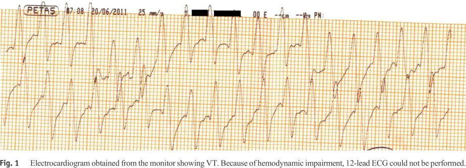
Click on images to make them larger. Image credit.[2] This is not the actual strip, but a strip of an adenosine pause edited to produce more asystole, which I have seen.
 

The side effect becomes much more of a problem when someone decides to treat the side effect, rather than wait for it to wear off.

We SLAM adenosine in because it wears off quickly. A minute, or two, of asystole is not a problem.

Giving a dose of epinephrine to a patient who had an SVT a minute ago and now has adenosine quickly wearing off – that may be a fatal problem.

But how effective is the Valsalva maneuver (VM)?
 

The VM is a non-invasive way to convert patients from SVT to sinus.It increases myocardial refractory period by increasing intrathoracic pressure thus stimulating baroreceptors in the aortic arch and carotid bodies Increases vagal tone (parasympathetic).[3]


Here is a big problem with the use of the Valsalva maneuver. It is just one method of attempting to stimulate the vagus nerve.

There are many other methods and they may be more successful. Carotid sinus massage (after auscultation for bruits), facial immersion in ice water (assuring that the airway does not become a problem), bearing down, blowing through a straw (even better may be a swizzle stick), digital circumferential sweep of the anus, coughing, . . . .

There are many ways of activating the vagal nerve, but my favorite is to act as if I have not started an IV before, go very s l o w l y with the insertion of the largest IV catheter I think I can get in the vein, and this has almost always broken the rhythm.

Yes, that is anecdotal, but I have only rarely needed to follow that with medication.

Yes, pain is not a nice thing, but it is much nicer than the side effects listed above.
 

Bottom Line: There is no standardized methods to perform a VM to terminate uncomplicated SVT that are evidence based.

Clinical Application: VM is a viable technique that is poorly researched for the conversion of SVT and should not be considered essential to attempt prior to chemical cardioversion.[2]


We need better vagal maneuvers.

We need good evidence on what works.
 

Go read the article and listen to the podcast.
 

Footnotes:

[1] ADENOCARD (adenosine) solution
[Astellas Pharma US, Inc.]

DailyMed
FDA Label

[2] Atrioventricular Re-entrant Tachycardia
Thumbnail Guide to Congenital Heart Disease
edited version of their adenosine ECG strip
Article

[3] SGEM#67: Shock the Monkey Tonight (Valsalva Maneuver for SVT)
Podcast Link: SGEM67
Date: March 23, 2014
Skeptics’ Guide to Emergency Medicine
Article and link to podcast

.

Who Needs a 12 Lead ECG?

ResearchBlogging.org
 

Do we do too many 12 lead ECGs on patients who do not have chest pain?

This is something that some people worry about.

Save the electrodes!

Those poor little electrodes are being abused!

Are electrodes being abused?
 

Women and the elderly with STEMI are particularly likely to present with atypical chief complaints such as dyspnea and weakness. Such patients experience significant delays in door-to-ECG time and treatment and have increased morbidity and mortality compared with patients who present with chest pain.5,9-12 [1]


Tiredness/weakness is the second best predictor of STEMI (ST segment Elevation Myocardial Infarction).

After chest pain (pressure, tightness, heaviness, squeezing, et cetera), the best predictor of STEMI is dyspnea in akll age ranges, but dyspnea indicates 20% of STEMIs in patients over 80 years old.

Are we helping anyone by avoiding 12 lead ECG (ElectroCardioGram) assessment?
 

Presenting chief complaints among 6,464 patients with STEMI. Chest pain decreased in frequency with age, whereas a chief complaint of dyspnea, weakness, syncope, or altered mental status all increased in frequency with age.[1]


Click on images to make them larger.


The advantage of a logarithmic chart is that there is greater distinction among the smaller numbers (such as the other complaints that make up less than 5% in the image above). The disadvantage is that large changes are flattened. I modified the dyspnea line to show how it would look on a linear scale (from 5% to 20%). As you can see, the ability to predict STEMI increases dramatically with age – more dramatically than the logarithmic scale suggests.


How should we remember all of this?

The authors came up with a nice simple flow chart (below).

This is for the ED, but is there a good reason for EMS to ignore these STEMIs?


Even in the 18-49 year old patients, dyspnea is about as likely to predict a STEMI as weakness is likely to predict a STEMI in an 80+ year old patient.

Chest pain still indicates about 50% of STEMI patients over 80, but we will miss half of STEMIs in this population if we only do 12 leads on chest pain patients.

Can an 80+ year old patient have a good quality of life after a STEMI?

Absolutely.

Also see When should you get an ECG? at Mill Hill Ave Command.

Footnotes:

[1] Development and validation of a prioritization rule for obtaining an immediate 12-lead electrocardiogram in the emergency department to identify ST-elevation myocardial infarction.
Glickman SW, Shofer FS, Wu MC, Scholer MJ, Ndubuizu A, Peterson ED, Granger CB, Cairns CB, Glickman LT.
Am Heart J. 2012 Mar;163(3):372-82. doi: 10.1016/j.ahj.2011.10.021.
PMID: 22424007 [PubMed – indexed for MEDLINE]

Glickman SW, Shofer FS, Wu MC, Scholer MJ, Ndubuizu A, Peterson ED, Granger CB, Cairns CB, & Glickman LT (2012). Development and validation of a prioritization rule for obtaining an immediate 12-lead electrocardiogram in the emergency department to identify ST-elevation myocardial infarction. American heart journal, 163 (3), 372-82 PMID: 22424007

.

A Recalled AED is Better Than No AED


Cardiac arrest. CPR in progress. Do not use the AED, because it has been recalled!

Wrong.


HeartStart automated external defibrillators from Philips Healthcare have been recalled.

What does the FDA (Food and Drug Administration) mean by recall?

Well, why was the recall issued?
 

Certain HeartStart automated external defibrillator (AED) devices made by Philips Medical Systems, a division of Philips Healthcare, may be unable to deliver needed defibrillator shock in a cardiac emergency situation, the U.S. Food and Drug Administration said today in a new safety communication for users of these previously recalled devices.[1]


A shock might not be delivered.

What does the FDA recommend?
 

“The FDA advises keeping all recalled HeartStart AEDs in service until you obtain a replacement from Philips Healthcare or another AED manufacturer, even if the device indicates it has detected an error during a self-test,” said Steve Silverman, director of the Office of Compliance in the FDA’s Center for Devices and Radiological Health.[1]


Do not take these AEDs out of service service until a replacement is present.

Why?

What about the lawyers?

But it’s defective!

Thinking is dangerous!
 

“Despite current manufacturing and performance problems, the FDA considers the benefits of attempting to use an AED in a cardiac arrest emergency greater than the risk of not attempting to use the defibrillator.”[1]


The benefit is greater than the risk.

There is risk with everything.

Anyone who tells you otherwise is selling something.

There is not benefit with everything.

Since the detection of an error during the self-test does not guarantee that the AED will not deliver a shock when needed, removing the AED without a replacement is more dangerous than leaving the AED in service.

These recalled AEDs are better than no AED.
 

Of course, if needed for use in an emergency, make every attempt to clear the error and use the device normally, as described in the Owner’s Manual.[2]


The manufacturer and the FDA agree that, in the case of these AEDs, something is better than nothing.

Are we really going to make a dead patient more dead by using a defective AED?

Footnotes:

[1] FDA issues safety communication on HeartStart automated external defibrillators from Philips Healthcare
FDA News Release
For Immediate Release: Dec. 3, 2013
Media Inquiries: Jennifer Rodriguez, 301-796-8232, jennifer.rodriguez@fda.hhs.gov
Consumer Inquiries: 888-INFO-FDA
News Release

[2] Philips HeartStart FRx and OnSite (HS1) automated external defibrillators (AEDs)
Phillips Healthcare
Maintenance Advisory

.

Is Earlier Better for Therapeutic Hypothermia? Part I

ResearchBlogging.org
 

When is the right time to begin TH (Therapeutic Hypothermia) to produce the best outcomes?

In the ICU (Intensive Care Unit)?

In the ED (Emergency Department)?

In the ambulance?

While the patient is still pulseless?

This question was asked in 2010.


Click on image to make it larger.
 

Favorable outcomes – 47.5% EMS TH vs 52.6% ED TH.

Worse outcome, but not statistically significant.

Discharge to home – 20.3% EMS TH vs 29.3% ED TH.

Worse outcome, but not statistically significant.

Discharge to rehabilitation – 27.1% EMS TH vs 23.3% ED TH.

Worse outcome, because these patients are not well enough to go home, but not statistically significant.

Dead – 52.5% EMS TH vs 46.6% ED TH.

Worse outcome, but not statistically significant.
 

The great tragedy of Science — the slaying of a beautiful hypothesis by an ugly fact. -Thomas Henry Huxley.
 

EMS TH was added to many EMS protocols because of a lack of clear evidence of harm. EMS needed to Just do something.

The results did not support EMS administration of chilled IV (IntraVenous) fluid for prehospital therapeutic hypothermia, but the study was stopped early, because –
 

At the interim analysis of the first 200 patients, the Steering Committee noted that there was no difference in the primary outcome measure and that it was extremely unlikely that such a difference would emerge between the groups. Therefore, the study was stopped because of futility after 234 patients had been enrolled.[1]


In other words – We will not let the numbers convince us that there is no benefit, because numbers that do not support a positive effect are futile?

If the data would have indicated a negative effect, but had not reached statistical significance, should we expect the Steering Committee to support continuing the study, or would they support discontinuing the study early to protect the enrolled patients, but leave the question unanswered?

When studies are discontinued early to protect patients, do they discourage further studies?

When studies are discontinued early to protect patients, do they only endanger future patients?

Or does early termination encourage further studies because there is not clear evidence of harm and we want to believe that our interventions are beneficial?
 

What if it works?

Most proposed treatments do not work, so this is just an excuse to continue using something dangerous. What if it works? is the logical fallacy that is used to justify harming patients with alternative medicine.

We should not harm vulnerable patients because of our unreasonable belief in wishful thinking.
 

If it helps just one patient it is worth it.

This is another logical fallacy, because it completely ignores the harm that the treatment causes.

Some patients will improve after almost any treatment – even cyanide.

That means that alternative medicine advocates could should endorse the use of cyanide, because if it helps just one patient . . . .

We need to have unbiased information about the real benefits (if any) and the real harms (if any), before we encourage using anything on vulnerable patients.
 

Is it good to just do something?

Or

Is it good to help patients?

If our responsibility is to help patients, one of the best ways to help patients is to avoid causing harm.

Just doing something, with no evidence of benefit, is causing harm.

How many EMS agencies have prehospital therapeutic hypothermia protocols because of a desire to just do something?

I have been criticized for not being a supporter of treatments that do not have evidence of benefit.

Am I a killjoy, desiring bad outcomes?

No.

I understand that treatment that does not have evidence of benefit is almost always going to do more harm than good.

Just do something?

No.

Just demand valid evidence of improved outcomes.

Footnotes:

[1] Induction of therapeutic hypothermia by paramedics after resuscitation from out-of-hospital ventricular fibrillation cardiac arrest: a randomized controlled trial.
Bernard SA, Smith K, Cameron P, Masci K, Taylor DM, Cooper DJ, Kelly AM, Silvester W; Rapid Infusion of Cold Hartmanns (RICH) Investigators.
Circulation. 2010 Aug 17;122(7):737-42. doi: 10.1161/CIRCULATIONAHA.109.906859. Epub 2010 Aug 2.
PMID: 20679551 [PubMed – indexed for MEDLINE]

Free Full Text from Circulation.

Bernard SA, Smith K, Cameron P, Masci K, Taylor DM, Cooper DJ, Kelly AM, Silvester W, & Rapid Infusion of Cold Hartmanns (RICH) Investigators (2010). Induction of therapeutic hypothermia by paramedics after resuscitation from out-of-hospital ventricular fibrillation cardiac arrest: a randomized controlled trial. Circulation, 122 (7), 737-42 PMID: 20679551

.

Why Did We Remove Atropine From ACLS? Part II


Continuing from Part I.

The AHA (American Heart Association) stopped recommendeding use of atropine for the treatment of PEA (Pulseless Electrical Activity) or asystole in the 2010 ACLS (Advanced Cardiac Life Support) guidelines.

There is not much information given, but that little bit of information just makes it more clear that we never had a good reason for making atropine a standard part of ACLS.

One sentence at a time, look at the reasoning –
 

Interventions Not Recommended for Routine Use During Cardiac Arrest
Atropine

Atropine sulfate reverses cholinergic-mediated decreases in heart rate and atrioventricular nodal conduction.[1]


There is a hypothetical justification for atropine based on physiology/pathophysiology.

There has been a hypothetical justification for every treatment found to be harmful. That hypothetical justification did not protect patients from real harm.
 

No prospective controlled clinical trials have examined the use of atropine in asystole or bradycardic PEA cardiac arrest.[2]


Why was a treatment that had never been demonstrated to improve outcomes recommended and the standard of care?

Without evidence of improved outcomes, should any treatment be used outside of controlled trials?
 

Lower-level clinical studies provide conflicting evidence of the benefit of routine use of atropine in cardiac arrest.34,295,–,304 [1]


To translate – Useless information is . . . useless.
 

There is no evidence that atropine has detrimental effects during bradycardic or asystolic cardiac arrest.[1]


Is atropine the alternative medicine of cardiac arrest?

This sentence contradicts the evidence review that led to the removal of atropine from the guidelines.

Here is a listing of the evidence that opposes the use of atropine for cardiac arrest.


Click on image to make it larger.[2]
 

While the evidence of harm is not great, the evidence of benefit is not great, either.

Evidence of worse outcomes from cardiac arrest is evidence of harm.

There are four studies – three that show a negative correlation with atropine and survival to discharge.

no evidence that atropine has detrimental effects?

The positive studies are also just showing correlation. Poor studies mean poor information. Why were we giving atropine based on poor information?

We were giving atropine based on wishful thinking.
 

Available evidence suggests that routine use of atropine during PEA or asystole is unlikely to have a therapeutic benefit (Class IIb, LOE B).[1]


We should not include treatments that do not have evidence of therapeutic benefit.
 

For this reason atropine has been removed from the cardiac arrest algorithm.[1]


For this reason, atropine should never have been included in the cardiac arrest algorithms.

For this reason, all treatments that do not have evidence of therapeutic benefit should have an expiration date.

If no evidence is provided, the treatment is removed from the guidelines.

This would apply to ventilations, epinephrine (Adrenaline), vasopressin (Pitressin), norepinephrine (Levophed), and phenylephrine (Neo-Synephrine) in cardiac arrest.

This would also apply to amiodarone (Cordarone), lidocaine (Xylocaine), and Magnesium in VF (Ventricular Fibrillation) cardiac arrest.
 

What does that leave us with?

Compressions in cardiac arrest.

Defibrillation in VF cardiac arrest.

Therapeutic hypothermia after resuscitation.

In Part III I will look at the most positive study supporting the use of atropine for cardiac arrest.

Footnotes:

[1] Atropine
2010 American Heart Association Guidelines for Cardiopulmonary Resuscitation and Emergency Cardiovascular Care Science
Part 8.2: Management of Cardiac Arrest
Interventions Not Recommended for Routine Use During Cardiac Arrest
Free Full Text from Circulation.

[2] Atropine for cardiac arrest
2010 American Heart Association Guidelines for Cardiopulmonary Resuscitation and Emergency Cardiovascular Care Science
Appendix: Evidence-Based Worksheets
Part 8 ALS
ALS-D-024B
Swee Han Lim
Evidence-Based Worksheet Download in PDF format.

That link is no longer available, but the overall page of evidence-based worksheets is available in PDF format here.

.

Epinephrine for V Tach – Instant Death or Effective Treatment?

ResearchBlogging.org
 

The patient has V Tach (Ventricular Tachycardia) with a pulse. After amiodarone is given the patient’s blood pressure drops and the patient becomes unstable. The patient is still awake, so cardioversion would be very painful and these physicians would need to get anesthesia to sedate the patient. I know – that anesthesia requirement is a bad policy and completely unnecessary for the safety of the patient, but it is politics in that facility. However, sedation for emergency cardioversion is very important.

There are other medications that might be effective at terminating V Tach.

What might happen if epinephrine is given?
 

Click on images to make them larger.
 

If take an ACLS (Advanced Cardiac Life Support) class, where the protocolized treatment of arrhythmias is taught, the instructor may tell you that you just killed the patient with epinephrine.

There were three patients treated with epinephrine for V Tach. They were not in the artificial environment of an ACLS class, and had not been taught by the epi for unstable V Tach = death ACLS instructors, so the patients did not know that they were supposed to die.

The physicians treating the V Tach patients had read the ACLS books, as well as a lot of other research on the treatment of arrythmias. The physicians chose to treat these patients with epinephrine.

47 year old male, 125/86 mmHg, heart rate of 170 with the rhythm above. 300 mg amiodarone over 5 min did not get rid of the V Tach, but it did appear to drop the blood pressure to 89/46 mmHg with profuse sweating, but without loss of consciousness. Perhaps that was also true of those taking care of the patient.

1 mg (1,000 μg) of 1:10,000 epinephrine given over less than 60 seconds was followed within 30 seconds by the changes below.


Heart rate increased to 180, briefly, blood pressure increased to 130/84 mmHg, then the rhythm converted.

Not to V Fib (Ventricular Fibrillation), but to a stable sinus tachycardia at a rate of 110.
 

The side effects of epinephrine were chest discomfort, nausea, and anxiousness.[1]


Well, he was only 47, so we got lucky with epinephrine at that young age.

How about a 64 year old male with a heart attack history, low ejection fraction and an AICD (Automated Implantable Cardioverter-Defibrillator), on bisoprolol 10 mg daily and amiodarone 200 mg daily for recurrent NSVT (Non-Sustained V Tach), conscious with a pressure of 85/50 mmHg and now a sustained V Tach at 140 beats/min?

A lower dose of epinephrine was given – 0.5 mg (500 μg) over less than 30 seconds was followed within 30 seconds by an increase of rate from 140 to 148, followed by conversion to a nice slow sinus rhythm.
 

The third patient had V Tach storm, which might seem even less likely to benefit from epinephrine.

A patient with a history of two heart attacks, taking carvedilol (12.5 mg twice daily) and amiodarone (200 mg daily) with an AICD, presenting with a pressure of 90/45 mmHg and a rate of 140+. He received six AICD shocks within 5 minutes in the hospital, but the rhythm returned to V Tach each time. 150 mg amiodarone was given over 15 min and the pressure dropped to 70/40 mmHg. Overdrive pacing was attempted with only a conversion of V Tach to V Fib, which was shocked by the AICD. Within 60 seconds, the V Tach was back. Blood pressure continued to fall to 65/30 mmHg.

Epinephrine was given – 0.5 mg (500 μg) over less than 30 seconds was followed within 30 seconds by an increase in blood pressure to 125/85 mmHg, followed by termination of V Tach within 90 seconds.
 

In cases of drug-resistant poorly tolerated VT, immediate external electrical cardioversion must be attempted. However, there are cases in which VT recurs immediately after the shock, and cardioversion involves the need for anesthesia when the patient is still conscious.[1]


Based on the cases reported herein, low doses of IV epinephrine may be able to terminate sustained monomorphic VT, when the arrhythmia is refractory to amiodarone used alone or in combination with beta-blockers and electrical cardioversion.[1]


I would not refer to 1,000 μg epinephrine, or even 500 μg, as low dose.

Dr. Scott Weingart discusses the use of epinephrine as a bolus dose pressor, but at much smaller doses.
 

Dose:

0.5-2 ml every 2-5 minutes (5-20 mcg)

No extravasation worries![2]


Do not use epinephrine for V Tach without discussing it with your medical director and obtaining permissions, assuming you work some place where the medical director has the authority. If you are a doctor, discuss this with cardiology before using it.

Read the discussion of the many possible confounders in the full text of the paper and learn a bit about cardiology and the ways that physiology misleads us.

Footnotes:

[1] Low doses of intravenous epinephrine for refractory sustained monomorphic ventricular tachycardia.
Bonny A, De Sisti A, Márquez MF, Megbemado R, Hidden-Lucet F, Fontaine G.
World J Cardiol. 2012 Oct 26;4(10):296-301. doi: 10.4330/wjc.v4.i10.296.
PMID: 23110246 [PubMed]

Free Full Text from PubMed Central.

[2] Push-Dose Pressors – Podcast 6
EMCrit
Dr. Scott Weingart
Article and podcast.

Bonny, A. (2012). Low doses of intravenous epinephrine for refractory sustained monomorphic ventricular tachycardia World Journal of Cardiology, 4 (10) DOI: 10.4330/wjc.v4.i10.296

.

Examples of Ventricular Tachycardia Caused by Amiodarone – Part I

ResearchBlogging.org
 

How much worse could the patient get if we give amiodarone?

But amiodarone doesn’t cause V Tach (Ventricular Tachycardia). Amiodarone stops V Tach.

Right?

If amiodarone can cause V Tach, shouldn’t someone have told us?


Click on image to make it larger.
 

This is a wide complex tachycardia, which should be presumed to be V Tach. The diagnosis of the rhythm as V Tach is supported by the P-wave dissociation and by the history of a heart attack a couple of weeks before. 19 QRS complexes in 5 seconds (multiply by 12 to get the rate for 60 seconds) = 228.

According to ACLS (Advanced Cardiac Life Support) what drug would be indicated for the above rhythm?

For the old timers, let’s throw in a bit of information that is supposed to be important in deciding among the various antiarrhythmics –
 

Transthoracic echocardiography was performed, and the ejection fraction was 34% based on the modified Simpson method.[1]


Does the ejection fraction matter in treating wide-complex tachycardia?
 

Note that amiodarone becomes the antiarrhythmic of choice (after failure of adenosine) if the patient’s cardiac function is impaired and the ejection fraction is <40% or there are signs of congestive heart failure.[2]


That was only mentioned in the 2000 ACLS guidelines. The 2005 ACLS guidelines and the 2010 ACLS guidelines do not mention ejection fraction in the treatment of V Tach.
 

Do we have good evidence that amiodarone is effective for stable V Tach?
 

Amiodarone has not been studied specifically for the pharmacological termination of hemodynamically stable VT, but it is effective in treating hemodynamically unstable VT and VF.78 81 82 83 84 85 86 87 88 89 90 91 [2]


That was written in 2000 and the answer was NO.

Now we have evidence that amiodarone may be effective in about one out of four patients – if we are prepared to wait a half hour, or more. In other words, it is rare for amiodarone to work and rarer still for amiodarone to work before we can get the patient to the hospital.[3],[4],[5]

We do have evidence that amiodarone may be a part of the long-term treatment of unstable V Tach. On the other hand, maybe not.

I do mean long-term treatment, even though these are unstable patients.
 

Twenty-seven (58.5%) of the 46 patients responded to intravenous amiodarone, and an additional 6 patients (13%) showed a late response to amiodarone (Fig. I). Fifteen of the 27 responders (33% of all patients) responded immediately (0 to 2 h), and 26 responded within 72 h . The other patient responded within 84 h (Fig. 2).[6]

 
Up to 84 hours to take effect.

Not exactly an emergency drug.

I have worked some very long shifts. I have never worked an 84 hour shift.

If amiodarone can take 84 hours to work, and the patient is receiving a variety of other treatments during those 84 hours, is it really the amiodarone that is working?

Compared with 84 hours, a less than two hour time to response seems almost instantaneous, but it is not fast for EMS.
 

This patient was treated with oral amiodarone for sustained V Tach during his previous hospital admission for an acute anterior MI (Myocardial Infarction) treated with a stent in the LAD (Left Anterior Descending) coronary artery.

This time he was treated with IV (IntraVenous) amiodarone. The problems began on the first day of IV amiodarone treatment.
 

On the first day of hospitalization, the patients had 10 attacks of pulseless VT treated with successful DC shocks. Occasionally, VT attacks degenerated to ventricular fibrillation.[1]


And continued –
 

On the second day of hospitalization, the patient had 14 pulseless VT attacks with successful DC shocks.[1]


But the V Tach stopped after the amiodarone was stopped and antithyroid therapy was begun.
 

Thyroid gland toxicity is one of the most important adverse effects of amiodarone and is called amiodarone-induced thyrotoxicosis. Thyrotoxicosis may alter arrhythmia and lead to frequent ventricular tachycardia attacks.[1]


All things are poison, and nothing is without poison; only the dose permits something not to be poisonous. – Paracelsus (1493-1541)
 

We do not seem to explain that well in EMS, or even in emergency medicine.
 

All antiarrhythmic agents have some degree of proarrhythmic effects.[7]


Amiodarone is one of the most dangerous drugs we carry in EMS.

How much thought do we give to the problems we can cause for our patients with this drug?

How much do we understand about the problems we could be causing when we push medications, such as amiodarone?

Footnotes:

[1] Therapy-resistant ventricular tachycardia caused by amiodarone-induced thyrotoxicosis: a case report of electrical storm.
Erdogan HI, Gul EE, Gok H, Nikus KC.
Am J Emerg Med. 2012 Nov;30(9):2092.e5-7. doi: 10.1016/j.ajem.2011.12.035. Epub 2012 Mar 3.
PMID: 22386340 [PubMed – indexed for MEDLINE]

[2] Amiodarone
2000 ECC Guidelines
Part 6: Advanced Cardiovascular Life Support
Section 5: Pharmacology I: Agents for Arrhythmias
Arrhythmias and the Drugs Used to Treat Them
Hemodynamically Stable Wide-/Broad-Complex Tachycardias
Treatment of Wide-Complex Tachycardias
Free Full Text from Circulation.

[3] Amiodarone is poorly effective for the acute termination of ventricular tachycardia.
Marill KA, deSouza IS, Nishijima DK, Stair TO, Setnik GS, Ruskin JN.
Ann Emerg Med. 2006 Mar;47(3):217-24. Epub 2005 Nov 21.
PMID: 16492484 [PubMed – indexed for MEDLINE]

[4] Amiodarone or procainamide for the termination of sustained stable ventricular tachycardia: an historical multicenter comparison.
Marill KA, deSouza IS, Nishijima DK, Senecal EL, Setnik GS, Stair TO, Ruskin JN, Ellinor PT.
Acad Emerg Med. 2010 Mar;17(3):297-306. doi: 10.1111/j.1553-2712.2010.00680.x.
PMID: 20370763 [PubMed – indexed for MEDLINE]

Free Full Text from Academic Emergency Medicine.

[5] Intravenous amiodarone for the pharmacological termination of haemodynamically-tolerated sustained ventricular tachycardia: is bolus dose amiodarone an appropriate first-line treatment?
Tomlinson DR, Cherian P, Betts TR, Bashir Y.
Emerg Med J. 2008 Jan;25(1):15-8.
PMID: 18156531 [PubMed – indexed for MEDLINE]

[6] Use of intravenous amiodarone for emergency treatment of life-threatening ventricular arrhythmias.
Helmy I, Herre JM, Gee G, Sharkey H, Malone P, Sauve MJ, Griffin JC, Scheinman MM.
J Am Coll Cardiol. 1988 Oct;12(4):1015-22.
PMID: 3417974 [PubMed – indexed for MEDLINE]

Page with link to Free Full Text Download in PDF format from J Am Coll Cardiol.

This is footnote 84 from the second 2000 ACLS quote.

[7] New Concerns From the International Guidelines 2000 Conference: Impaired Hearts and “Proarrhythmic Antiarrhythmics”
2000 ECC Guidelines
Part 6: Advanced Cardiovascular Life Support
Section 5: Pharmacology I: Agents for Arrhythmias
Arrhythmias and the Drugs Used to Treat Them
Hemodynamically Stable Wide-/Broad-Complex Tachycardias
Free Full Text from Circulation.

Erdogan, H., Gul, E., Gok, H., & Nikus, K. (2012). Therapy-resistant ventricular tachycardia caused by amiodarone-induced thyrotoxicosis: a case report of electrical storm The American Journal of Emergency Medicine, 30 (9), 209200000-2147483647 DOI: 10.1016/j.ajem.2011.12.035

Marill, K., deSouza, I., Nishijima, D., Stair, T., Setnik, G., & Ruskin, J. (2006). Amiodarone Is Poorly Effective for the Acute Termination of Ventricular Tachycardia Annals of Emergency Medicine, 47 (3), 217-224 DOI: 10.1016/j.annemergmed.2005.08.022

Marill, K., deSouza, I., Nishijima, D., Senecal, E., Setnik, G., Stair, T., Ruskin, J., & Ellinor, P. (2010). Amiodarone or Procainamide for the Termination of Sustained Stable Ventricular Tachycardia: An Historical Multicenter Comparison Academic Emergency Medicine, 17 (3), 297-306 DOI: 10.1111/j.1553-2712.2010.00680.x

Tomlinson, D., Cherian, P., Betts, T., & Bashir, Y. (2008). Intravenous amiodarone for the pharmacological termination of haemodynamically-tolerated sustained ventricular tachycardia: is bolus dose amiodarone an appropriate first-line treatment? Emergency Medicine Journal, 25 (1), 15-18 DOI: 10.1136/emj.2007.051086

Helmy, I., Herre, J., Gee, G., Sharkey, H., Malone, P., Sauve, M., Griffin, J., & Scheinman, M. (1988). Use of intravenous amiodarone for emergency treatment of life-threatening ventricular arrhythmias Journal of the American College of Cardiology, 12 (4), 1015-1022 DOI: 10.1016/0735-1097(88)90470-6

.

What Does a Moon Landing ECG Look Like?


Some interesting ECG from the first people to land on the Moon.

During landing, they were running low on fuel and Buzz Aldrin appears to be showing signs of stress.


Click on images to make them larger.
 

The image above is from the following video describing the monitoring of the ECGs of the astronauts.


The reason this is in the news now is that one of four of the ECGs of Neil Armstrong as he stepped on the Moon was scheduled to be auctioned, but has been pulled due to questions about whether the owners have clear title to these items.[1]

How low on fuel were they? Were they going to crash?

They were just going to have to abort the mission and return to the Command Module, but after years of preparation for a Moon landing, that can be a huge amount of stress.


“EKG Recordings Taken as Apollo 11 Commander Neil Armstrong Took Man’s First Step on the Moon” and “4:13:24:28 Ground Elapsed Time.” Sheet is signed and inscribed in pencil, “To Paul Jones, The heartbeats that made this accomplishment possible as recorded at MCC on my console. Keep up your heart work. Charles A. Berry M.D.” Presentation also bears a Neil Armstrong autopen signature. Sheet is matted and framed with mission patches from Apollo 7, Apollo 8, Apollo 9, Apollo 10, Apollo 11, Apollo 12, Apollo 13, and two Snoopy patches, to an overall size of 20.75 x 24.75.[2]


Look at Buzz Aldrin’s ECG. The rate is about 400 BPM (Beats Per Minute).

Can a human heart beat that fast?

I have seen close to 300 BPM in a febrile infant.

Is the following rate possible for a human?


The rate is probably not possible.

The reason it looks so fast is most likely because the paper is being fed at a much slower speed than usual.

Conversely, we can get a better idea of what a very fast tachycardia looks like by speeding up the paper feed rate from the standard 25 mm/second to 50 mm/second or by manually pulling the paper through the printer faster than its normal rate.
 

EKG strip, six inches long, taken as Apollo 11 Commander Neil Armstrong took man’s first step on the moon. This is an actual strip of the EKG from Armstrong’s heart monitor at the moment he stepped onto the lunar surface.[2]


Compare that with a “six second section” of Buzz Aldrin’s ECG during their very low on fuel landing.


At 12.5 mm/second, this would be a rate of about 200 BPM, faster than the calculated maximum heart rate, but still capable of being a sinus tachycardia that is only associated with minor/moderate symptoms. At 10 mm/second, this would be a rate of about 160 BPM, which I regularly exceed (and recover from without any need for adenosine or cardioversion).
 

[youtube]sTBIr65cL_E[/youtube]
 

Several times you hear them checking with the flight surgeon and receiving a “Go,” each time. A heart rate of 400 should have resulted in something other than a “Go.” A few questions to Buzz Aldrin about how he is feeling would have been prudent.

If I have a patient with a heart rate of 400 and I do not ask a few questions about how the patient is doing, it would probably be because the patient is not capable of communicating. How are you feeling, hummingbird?
 

In general, sinus tachycardia is a response to other factors and, thus, it rarely (if ever) is the cause of instability in and of itself.[3]


At EMS 12 Lead, there is an excellent discussion of sinus tachycardia, and the nonsense of assuming that anything faster than 150 BPM is an SVT that needs adenosine or cardiversion.[4] This includes comments from Dr. John Mandrola and Dr. Mark Perrin.
 

Go read it.
 

Footnotes:

[1] Neil Armstrong’s ‘Heartbeat,’ Apollo Joystick Pulled from Auction
by Robert Z. Pearlman, collectSPACE.com Editor
Date: 20 May 2013 Time: 04:34 PM ET
Article

[2] Neil Armstrong’s Heartbeat – EKG Up For Auction
By Patrick Lockerby
May 5th 2013 04:20 PM
Science 2.0
Article

[3] Overview
2010 American Heart Association Guidelines for Cardiopulmonary Resuscitation and Emergency Cardiovascular Care Science
Part 8: Adult Advanced Cardiovascular Life Support
Part 8.3: Management of Symptomatic Bradycardia and Tachycardia
Free Full Text from Circulation.

[4] The Trouble With sinus Tachycardia
April 30, 2013
David Baumrind
EMS 12 Lead
Article

.