Without evidence of benefit, an intervention should not be presumed to be beneficial or safe.

- Rogue Medic

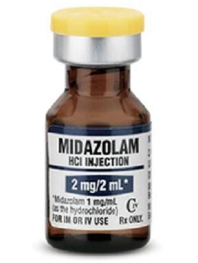
Intramuscular Midazolam for Seizures – Part III


ResearchBlogging.org
Also posted over at Paramedicine 101 (now at EMS Blogs) and at Research Blogging. Go check out the excellent material at these sites.

I have already pointed out my disappointment with the references of this large double-blind, randomized, noninferiority trial comparing IM (IntraMuscular) midazolam (Versed) with IV (IntraVenous) lorazepam (Ativan). One of those criticisms appears to be just due to a typographical error. The footnote in the text was 11, but the footnote should have been 1.
 

The relationships among benzodiazepine dose, respiratory depression, and subsequent need for endotracheal intubation are poorly characterized, but higher doses of benzodiazepines may actually reduce the number of airway interventions. Our data are consistent with the finding that endotracheal intubation is more commonly a sequela of continued seizures than it is an adverse effect of sedation from benzodiazepines.11 [1]

Here is some of the information from footnote 1. One interesting aspect of this double-blind study is that there is a placebo group. Patients received 2 mg IV lorazepam, 5 mg IV diazepam (Valium), or IV placebo. Treatment could be repeated one time if seizures continued for more than 4 minutes or if seizures recurred.
 

Cardiorespiratory complications before arrival at the hospital and at the time of transfer were important secondary outcomes that relate to the safety of out-of-hospital therapy with intravenous benzodiazepines. Despite concern regarding the adverse effects of these agents, we found a trend toward lower rates of out-of-hospital complications (primarily respiratory compromise) in the active-treatment groups than in the placebo group. This suggests that respiratory complications associated with prolonged seizures may be more pronounced than those caused by intravenous lorazepam and diazepam given at relatively low doses.[2]


The doses are low. The lorazepam dose is only half of the 4 mg used in the IV lorazepam vs. IM midazolam study.
 

The doses of midazolam and lorazepam used in this trial are consistent with the most effective doses for the treatment of status epilepticus that are reported in the literature.9,10 Although these initial doses are higher than the ones used by many EMS systems and emergency physicians, they are the same as those approved for this indication and are in line with those used by epileptologists.[1]

Is there added safety from the lower doses?

The epilepsy specialists and the FDA (Food and Drug Administration) do not recommend lower doses.

Were the low doses effective?

2 mg midazolam?

Does anyone really expect such a small dose to make a difference?

Despite the beneficial outcomes associated with intravenous lorazepam and diazepam, 41 to 57 percent of patients who received active treatment were still in status epilepticus at the time of arrival at the emergency department. These patients were more than twice as likely to require intensive medical care as those whose seizures ended outside the hospital. Differences in the causes of the episodes of status epilepticus are unlikely to account for this difference. These observations, coupled with the favorable risk–benefit profile associated with lorazepam and diazepam in this trial, suggest that higher doses should be studied to define the optimal therapy for patients with out-of-hospital status epilepticus.[2]


An editorial refers to the study just published[1] and to the benzodiazepine vs. placebo study.[2] Describing the complications in the placebo study, the author wrote –
 

Successful termination was much more common in the two groups that received benzodiazepines (59% with lorazepam, 43% with diazepam, and 21% with placebo). Since respiratory distress was twice as common in the group given placebo as in either of the groups given a benzodiazepine, the best way to avoid the need for intubation is to stop seizure activity.[3]


This presents an interesting conundrum. Doses of benzodiazepines (midazolam, lorazepam, diazepam, . . .) are often limited, due to a fear of causing respiratory complications.

When treating seizures, higher doses of benzodiazepines may actually protect patients from respiratory complications.

With a fatality rate around 10%, seizures are certainly not benign.

Maybe early treatment with high dose benzodiazepines can significantly decrease that fatality rate.

Finally, relatively few out-of-hospital interventions have been evaluated in randomized controlled trials,16 and when they have been evaluated carefully, therapies with intuitive appeal have often been found either to lack benefit or to cause harm to patients.17-20 [2]


The irony is that we may be doing the opposite by limiting doses of benzodiazepines to less than what is recommended by the FDA.

What do you think?

I have written about this in Intramuscular Midazolam for Seizures – Part I,
Part II,
Part III,
Part IV,
Part V,
Part VI,
Misrepresenting Current Topics in EMS Research from EMS Expo – RAMPART,
and Images from Gathering of Eagles Presentation on RAMPART.

Footnotes:

[1] Intramuscular versus intravenous therapy for prehospital status epilepticus.
Silbergleit R, Durkalski V, Lowenstein D, Conwit R, Pancioli A, Palesch Y, Barsan W; NETT Investigators.
N Engl J Med. 2012 Feb 16;366(7):591-600.
PMID: 22335736

Free Full Text from N Engl J Med.

[2] A comparison of lorazepam, diazepam, and placebo for the treatment of out-of-hospital status epilepticus.
Alldredge BK, Gelb AM, Isaacs SM, Corry MD, Allen F, Ulrich S, Gottwald MD, O’Neil N, Neuhaus JM, Segal MR, Lowenstein DH.
N Engl J Med. 2001 Aug 30;345(9):631-7. Erratum in: N Engl J Med 2001 Dec 20;345(25):1860.
PMID: 11547716 [PubMed – indexed for MEDLINE]

Free Full Text from N Engl J Med. with link to PDF Download

[3] Intramuscular versus intravenous benzodiazepines for prehospital treatment of status epilepticus.
Hirsch LJ.
N Engl J Med. 2012 Feb 16;366(7):659-60. No abstract available.
PMID: 22335744 [PubMed – in process]

Silbergleit, R., Durkalski, V., Lowenstein, D., Conwit, R., Pancioli, A., Palesch, Y., & Barsan, W. (2012). Intramuscular versus Intravenous Therapy for Prehospital Status Epilepticus New England Journal of Medicine, 366 (7), 591-600 DOI: 10.1056/NEJMoa1107494

Alldredge BK, Gelb AM, Isaacs SM, Corry MD, Allen F, Ulrich S, Gottwald MD, O’Neil N, Neuhaus JM, Segal MR, & Lowenstein DH (2001). A comparison of lorazepam, diazepam, and placebo for the treatment of out-of-hospital status epilepticus. The New England journal of medicine, 345 (9), 631-7 PMID: 11547716

Hirsch LJ (2012). Intramuscular versus intravenous benzodiazepines for prehospital treatment of status epilepticus. The New England journal of medicine, 366 (7), 659-60 PMID: 22335744

.
Nifekalant versus lidocaine for in-hospital shock-resistant ventricular fibrillation or tachycardia


ResearchBlogging.org

Also posted over at Paramedicine 101 (now at EMS Blogs) and at Research Blogging. Go check out the excellent material at these sites.

This is an interesting study for several reasons. One is the ability of the authors to act out parts of Through the Looking-Glass.[1] VF and VT are Ventricular Tachycardia and Ventricular Fibrillation.

If life-threatening VF or VT persists despite repeated defibrillation shocks, an additional antiarrhythmic drug is required.[2]

The next paragraph points out that there is no requirement according to ACLS.

The American Heart Association guideline for advanced cardiac life support (ACLS) states that when VF/pulseless VT persists after two to three shocks plus CPR and administration of a vasopressor, the physician should consider administering an anti-arrhythmic such as amiodarone, and lidocaine may be considered if amiodarone is unavailable.1 [2]

Believing that should consider administering is the same as an additional antiarrhythmic drug is required requires the same lack of illogic as used by the Queen, when instructing Alice to practice believing impossible things.

Then there are the obvious questions. Why compare nikefelant with lidocaine? Why not compare nikefelant with amiodarone? Why not compare nikefelant with an antiarrhythmic that is more effective than amiodarone – procainamide, sotalol, or ajmaline?

Lidocaine is probably used because the IRB (Institutional Review Board) would consider it unethical to have a placebo group. Lidocaine is the placebo, but with less safety than the placebo.

It was by comparing amiodarone with lidocaine that ACLS ended up including amiodarone for VT/VF (Ventricular Tachycardia/Ventricular Fibrillation) cardiac arrests. That was a huge boon for Wyeth. We were told that the improved survival to admission was important. We were told that survival studies – the only studies that matter in resuscitation – were being done. We have had only silence since then.

We should conclude that amiodarone does not improve survival.

No. That is not the right conclusion. If amiodarone produced survival as good as placebo, then that study would have been published and used to justify giving amiodarone. At least we are doing something! That is what people want to believe in.

The only reasonable conclusion is that Wyeth did not publish the results because the survival in the amiodarone group was significantly worse than in the placebo group, or Wyeth stopped the study early, because it was trending toward statistically significant harm from amiodarone.

If the study never reaches statistical significance, they can always rely on there being no proof of harm. That is what we have now and there are plenty of people claiming that no proof of harm means obvious benefit.


Original cartoon

We have a lot of ignorant/willfully ignorant people encouraging us to just give drugs because we cannot prove that these drugs are harmful. We cannot provide evidence that the drugs are harmful, because it is almost impossible to approve a placebo-controlled study to find out. The IRBs claim that it would be unethical to deprive patients of the Standard Of Care, no matter how harmful that Standard Of Care may be. If the IRBs approve a study, the politicians oppose the study.[3]

There were some interesting differences between the lidocaine and nikefalant groups.

The number of shocks before study-drug administration did not differ between the two arms, although epinephrine use before study-drug administration was significantly higher in the lidocaine arm (Table 2).[2]

Epinephrine use
Nikefalant   6 out of 27 = 22.2%
Lidocaine   20 out of 28 = 71.4%
With a p value of <0.001

According to ACLS, an antiarrhythmic should not be considered until after a pressor is considered.

Patients with nifekalant were more likely to have ROSC compared with patients with lidocaine (Table 3). However, there was no difference in 1-month survival or survival to hospital discharge between the nifekalant arm and the lidocaine arm.[2]

The overall outcome, such as survival rate, of patients with shock-resistant VF or VT is poor regardless of the pharmacological intervention. Nevertheless, termination of VF or VT and recovery of ROSC by nifekalant is important in the initial stage of resuscitation, because we cannot rescue the patients unless VF or VT is converted.[2]

They assume that the VF/VT will not be converted without a drug.

They assume that converting more VF/VT will lead to more survival even though there continues to be absolutely no evidence to support this hope.

It is reasonable to assume that the short-term thrill of conversion of VF/VT to a better rhythm comes at the expense of long-term harm to the patient.

We need to stop falling for feel good endpoints that encourage us to harm our patients.

If others are NOT helping their patients with these drugs, we want to be NOT helping our patients, too!

Footnotes:

[1] Through the Looking-Glass
by Lewis Carroll
The Millennium Fulcrum Edition 1.7
CHAPTER V. Wool and Water

‘I can’t believe THAT!’ said Alice.

‘Can’t you?’ the Queen said in a pitying tone. ‘Try again: draw a long breath, and shut your eyes.’

Alice laughed. ‘There’s no use trying,’ she said: ‘one CAN’T believe impossible things.’

‘I daresay you haven’t had much practice,’ said the Queen. ‘When I was your age, I always did it for half-an-hour a day. Why, sometimes I’ve believed as many as six impossible things before breakfast.

[2] Nifekalant versus lidocaine for in-hospital shock-resistant ventricular fibrillation or tachycardia.
Shiga T, Tanaka K, Kato R, Amino M, Matsudo Y, Honda T, Sagara K, Takahashi A, Katoh T, Urashima M, Ogawa S, Takano T, Kasanuki H; Refractory VT/VF, Prospective Evaluation to Differentiate Lidocaine Efficacy from Nifekalant (RELIEF) Study Investigators.
Resuscitation. 2010 Jan;81(1):47-52. Epub 2009 Nov 13.
PMID: 19913983 [PubMed – indexed for MEDLINE]

[3] Effect of adrenaline on survival in out-of-hospital cardiac arrest: A randomised double-blind placebo-controlled trial
Jacobs IG, Finn JC, Jelinek GA, Oxer HF, Thompson PL.
Resuscitation. 2011 Sep;82(9):1138-43. doi: 10.1016/j.resuscitation.2011.06.029. Epub 2011 Jul 2.
PMID: 21745533

Free Full Text PDF Download from semanticscholar.org
 

This study was designed as a multicentre trial involving five ambulance services in Australia and New Zealand and was accordingly powered to detect clinically important treatment effects. Despite having obtained approvals for the study from Institutional Ethics Committees, Crown Law and Guardianship Boards, the concerns of being involved in a trial in which the unproven “standard of care” was being withheld prevented four of the five ambulance services from participating.

In addition adverse press reports questioning the ethics of conducting this trial, which subsequently led to the involvement of politicians, further heightened these concerns. Despite the clearly demonstrated existence of clinical equipoise for adrenaline in cardiac arrest it remained impossible to change the decision not to participate.

Shiga, T., Tanaka, K., Kato, R., Amino, M., Matsudo, Y., Honda, T., Sagara, K., Takahashi, A., Katoh, T., Urashima, M., Ogawa, S., Takano, T., & Kasanuki, H. (2010). Nifekalant versus lidocaine for in-hospital shock-resistant ventricular fibrillation or tachycardia Resuscitation, 81 (1), 47-52 DOI: 10.1016/j.resuscitation.2009.09.027

Jacobs IG, Finn JC, Jelinek GA, Oxer HF, & Thompson PL (2011). Effect of adrenaline on survival in out-of-hospital cardiac arrest: A randomised double-blind placebo-controlled trial. Resuscitation, 82 (9), 1138-43 PMID: 21745533

.

Charging the Defibrillator While Continuing Chest Compressions – Part II


ResearchBlogging.org
Also posted over at Paramedicine 101 (now at EMS Blogs) and at Research Blogging. Go check out the excellent material at these sites.

Continuing, after a 6 month delay, a discussion of an EMS 12 Lead article from Part I. ACLS (Advanced Cardiac Life Support) recommends charging the defibrillator during compressions. This is no less of a recommendation than giving epinephrine. How many people ignore ACLS guidelines for compressions during charging, but claim that it is evil to disobey anything ACLS recommends on epinephrine, amiodarone, or ventilations?

Analyses of VF waveform characteristics predictive of shock success have documented that the shorter the time interval between the last chest compression and shock delivery, the more likely the shock will be successful.141 A reduction of even a few seconds in the interval from pausing compressions to shock delivery can increase the probability of shock success.142 [1]

Extra pauses in compressions add to the time without compressions.

If the medic/nurse/doctor using a manual defibrillator recognizes a shockable rhythm, why not provide compressions while charging the defibrillator?

Some people will say that this is dangerous.


Image credit.

But if someone accidentally delivers a shock during compressions, people will be killed!

In a systematic review, Hoke et al. summarized 29 reports of accidental defibrillator discharges, of which only 15 occurred during resuscitation attempts.21 Symptoms included tingling sensations, discomfort, and minor burns, but no long term effects or major consequences were reported.[2]

Where are the dead bodies we hear so much about?

Where are the medics/nurses/doctors needing to be defibrillated back to life?

There was only one incident where a shock was delivered while a rescuer was actively performing chest compressions. However, the compression transcript continued without any visible change to CPR administration, suggesting that the rescuer was unaffected by the event. Review of clinical records and audio transcripts revealed no evidence of inadvertent shocks to rescuers. In addition, there was no significant difference in the incidence of inappropriate shocks to patients associated with charging during compressions (20.0% vs 20.1%; p = 0.97). [2]

In this study, there was one case of a shock being delivered during compressions, but nobody seems to have been affected by this shock.

What happened to the automatic death that ACLS instructors spend so much time describing?

Where is the evidence?

In the current study, charging during compressions decreased median pre-shock pause by over 10 s, which previous studies suggest could have a dramatic effect on clinical outcomes. We previously reported an almost two-fold increase in the chances of successful defibrillation for every 5 s reduction in the pre-shock pause.9 Similarly, Eftestøl et al. found that a 10 s hands-off period prior to defibrillation would roughly halve the probability of obtaining ROSC.6 [2]

The risk to rescuers appears to be minimal, but the possible benefit to patients may be dramatic.


Click on image to make it larger.

The difference in time without compressions is significant.

Interestingly, we found that the most efficient technique with regard to minimizing pauses was not the AHA recommended method of pausing to analyze, resuming CPR to charge, and then pausing again to defibrillate. Rather, charging at the end of every 2 min CPR cycle in anticipation of a shockable rhythm and then pausing only once, briefly, to both analyze and either shock or disarm was associated with significantly shorter total pause duration in the 30 s preceding defibrillation. [2]

If we see asystole, we do not deliver a shock. We cancel the shock.

If we see PEA (Pulseless Electrical Activity, such as sinus rhythm, sinus tachycardia, sinus bradycardia, or any other non-shockable rhythm), we do not deliver a shock. We cancel the shock.

Cancelling the shock is not going to be the same for each defibrillator, but we do need to know how to cancel the shock for each machine we use. We can read the instructions.

How?

We can turn on the monitor, charge it up to the setting we would use to defibrillate, and try to figure out ways to get the charged defibrillator to turn the shock off. We should already know how to do this.

All that appears to be required is competence. Why is that so difficult?

Why do we keep making excuses for misbehavior?

Footnotes:

[1] CPR Before Defibrillation
2010 American Heart Association Guidelines for Cardiopulmonary Resuscitation and Emergency Cardiovascular Care Science
Part 8: Adult Advanced Cardiovascular Life Support
Rhythm-Based Management of Cardiac Arrest
Defibrillation Strategies
Free Full Text from Circulation with links to Free Full Text PDF

[2] Safety and efficacy of defibrillator charging during ongoing chest compressions: a multi-center study.
Edelson DP, Robertson-Dick BJ, Yuen TC, Eilevstjønn J, Walsh D, Bareis CJ, Vanden Hoek TL, Abella BS.
Resuscitation. 2010 Nov;81(11):1521-6.
PMID: 20807672 [PubMed – indexed for MEDLINE]

Edelson, D., Robertson-Dick, B., Yuen, T., Eilevstjønn, J., Walsh, D., Bareis, C., Vanden Hoek, T., & Abella, B. (2010). Safety and efficacy of defibrillator charging during ongoing chest compressions: A multi-center study Resuscitation, 81 (11), 1521-1526 DOI: 10.1016/j.resuscitation.2010.07.014

.

Intraosseous Versus Intravenous Vascular Access During Out-of- Hospital Cardiac Arrest – A Randomized Controlled Trial

ResearchBlogging.org

Also posted over at Paramedicine 101 (now at EMS Blogs) and at Research Blogging. Go check out the excellent material at these sites.

For treatment of medical cardiac arrest patients, which is better – IO (IntraOsseous) or IV (IntraVenous) access for medication administration?

Since no medications have ever been demonstrated to improve survival from cardiac arrest (only chest compressions and defibrillation have), the most important consideration will be what method results in the least interruption of compressions and the least interference with defibrillation.

All patients eligible for inclusion in this study had their first attempt at vascular access randomized to one of 3 locations: proximal tibial intraosseous, proximal humeral intraosseous, or peripheral intravenous. The proximal tibial insertion site was located medial to the tibial tuberosity, or just below the patella along the flat aspect of the tibia. The proximal humerus insertion site was defined as the greater tubercle of the anterior humeral head 1 cm proximal to the surgical neck of the humerus. Peripheral intravenous catheter placement could occur at any accessible peripheral vein but preferably at the antecubital fossa; the external jugular vein was not an option provided for catheterization.[1]


Proximal tibial access points.


Proximal humeral access point.
Images credit.

Does this hurt? No. The patients are unresponsive and pulseless (dead), but even live patients and EMS personnel (who have tried this on themselves) report very little pain.

Overall success took into account a failure to maintain initial vascular access during the course of resuscitation, which included needle dislodgement or the inability to successfully administer medications or fluid at any time during the resuscitation.[1]

Those would interfere with the one claimed benefit – ability to deliver medication.

There was no difference in time to success for either of the intraosseous routes compared with the peripheral intravenous route.[1]


Click on images to make them larger.

The time to success is interesting. The times for the humeral site are similar to the tibial IO and the IV for placement and first drug administration – at least at the low end of the IQRs (InterQuartile Ranges). The problem is that the upper end of the IQRs is much longer than for the other methods. This is in part due to the low number of patients, which is partially explained by the 13 protocol violations – all in favor of the tibial IO site. The lack of familiarity of paramedics probably also contributes, resulting in much longer times for some of the paramedics.

Finally, there were 13 protocol violations that favored the tibial intraosseous route, which may have been an indicator of bias among paramedics for that route and therefore could have resulted in confounding of the study results.[1]

It may be that this group of paramedics was much more comfortable with the tibial IO, than with the humeral IO and this led to a greater likelihood of coming up with excuses for protocol violations. This may also have led to the performance differences. I have seen similar differences with the introduction of a new type of IV catheter to some services. There can be a lot of conscious and unconscious resistance to the new method, but after some familiarity develops, things tend to return to normal.

In the literature, intraosseous needle insertions have been linked to local wound infections, osteomyelitis, fat emboli, and compartment syndrome.18-20 During this study, there was no mechanism in place for EMS or hospital personnel to report complications in the use of the intraosseous device.[1]

That would be good to know, but this was not one of the goals of the study.

The average weight of patients in the humeral intraosseous group was greater than that of individuals in either of the other 2 arms of the study. This increased weight may have been associated with a difficulty in obtaining or maintaining vascular access.[1]

Weight can be a problem for any method of IV/IO access, so this is a very important limitation.

Weight – mean (SD)

Overall – 97.3 kg (2.7)

Humeral IO – 103.9 kg (6.5)

IV – 97.7 kg (3.8)

Tibial IO – 91.5 kg (3.9)

An average weight of 228.6 pounds (103.9 kg) in the humeral IO group, but only 201.3 pounds (91.5 kg) in the tibial IO group? 27.3 pounds difference (13.8% difference).

That strongly suggests a problem.

The proximal humerus can also prove tenuous during cardiac arrest because it is centered near the upper torso, where resuscitation efforts are occurring, including airway management, ongoing chest compressions, and rescuer interchange. The constant activity creates a tremendous amount of movement and further increases the risk of unintentional needle dislodgement, which was verified during the debriefing session after each out-of-hospital cardiac arrest, with paramedics frequently citing entanglement of the humeral intraosseous line, leading to dislodgement.[1]

The peripheral intravenous site is the most commonly used vascular access by all health care providers, yet it proved successful in less than 50% of cases in this study.[1]

No matter how bad the success of the humeral IO was, the IV success was even worse – less than 50% first attempt success.

Do IOs improve outcomes?

IOs may make it less likely that compressions will be interrupted, but we cannot tell from this study.

IOs may make it more likely that potentially harmful medications will be given.

The most interesting numbers I saw were the total fluid infused – twice as much in the IV group as in either IO group. No explanation is given, other than the possible slower flow rate for an IO. This may help to prevent fluid overload for those patients not in need of having an IV line accidentally left wide open.

There is no evidence that IOs, IVs, tubes, or medications improve survival to discharge with a working brain.

ACLS drug therapy during CPR is often associated with increased rates of ROSC and hospital admission but not increased rates of long-term survival with good neurologic outcome.[2]

Will this make the Three Stooges Pit Crew concept less of a comedy of errors to implement?

Probably not, but we can hope that the AHA does the right thing and eliminates all of the treatments that don’t work – ventilation, intubation, IV access, IO access, epinephrine, amiodarone, lidocaine, atropine – wait, they actually did remove atropine, so there is hope.

Footnotes:

[1] Intraosseous Versus Intravenous Vascular Access During Out-of-Hospital Cardiac Arrest: A Randomized Controlled Trial.
Reades R, Studnek JR, Vandeventer S, Garrett J.
Ann Emerg Med. 2011 Dec;58(6):509-16.
PMID: 21856044 [PubMed – in process]

[2] Medications for Arrest Rhythms
Part 8: Adult Advanced Cardiovascular Life Support
2010 American Heart Association Guidelines for Cardiopulmonary Resuscitation and Emergency Cardiovascular Care
Part 8.2: Management of Cardiac Arrest
Free Full Text Article with links to Free Full Text PDF download

Reades R, Studnek JR, Vandeventer S, & Garrett J (2011). Intraosseous Versus Intravenous Vascular Access During Out-of-Hospital Cardiac Arrest: A Randomized Controlled Trial. Annals of emergency medicine, 58 (6), 509-16 PMID: 21856044

.

Does Epinephrine Improve Survival from Cardiac Arrest


ResearchBlogging.org
Also posted over at Paramedicine 101 (now at EMS Blogs) and at Research Blogging. Go check out the excellent material at these sites.

Even though epinephrine (adrenaline) is used automatically in cardiac arrest, and there is evidence that epinephrine helps to produce a pulse (ROSC – Return Of Spontaneous Circulation), there is no evidence that epinephrine improves the only survival statistic that matters – discharge from the hospital with a brain that still works. There were so many deviations from assignment protocol in their 2009 study,[1] that the authors decided to examine the results based on what treatment patients actually received. They refer to epinephrine as adrenaline, which is the same drug. I will use adrenaline for consistency.

Our randomized study was analyzed on an intention-to-treat basis.4 As expected; some patients in the intravenous group had achieved ROSC before adrenaline could be given, while some in the no-intravenous group received adrenaline for different reasons. For example, it was permitted to place the IV line 5 min after ROSC. If re-arrest occurred, adrenaline could be administered if indicated by the CPR guidelines.7 [2]

In the no andrenaline group, 37 of the 433 patients did receive andrenaline.

In the adrenaline group, 85 of the 418 patients did not receive andrenaline.

For 3 patients, the authors were unable to tell whether andrenaline was given and these patients were excluded.

This changes the data to 367 patients in the adrenaline group and 481 patients in the no adrenaline group.

Patients in the adrenaline group were more likely to be admitted to hospital and an intensive care unit compared to the no-adrenaline group (OR 2.5 CI 1.9, 3.4 and OR 1.4 CI 1.0, 1.9, respectively). [2]

This is nothing new. Patients receiving andrenaline are more likely to have ROSC. All that really matters is what happens after ROSC.

If the patient loses pulses after ROSC, giving more adrenaline may not produce the desired effect – another ROSC.

First look at Table 1. The duration of CPR is much longer with the adrenaline group. Is this because of patients losing pulses?


Click on images to make them larger.

You can also see how few drugs were given to the no adrenaline group. They were not supposed to receive any drugs, but the use of adrenaline was the only criterion for reassigning patients in this reanalysis of the data. Atropine was given to 2% of the no adrenaline group and amiodarone was given to 2%. Was there overlap of these patients? We can’t tell.

The defibrillations were also significantly different. More patients were shocked in the adrenaline group, but more patients in the adrenaline group were in VF (Ventricular Fibrillation) initially. How many of the patients with PEA (Pulseless Electrical Activity) or Asystole developed VF after adrenaline? More shocks were also used for each patient. Was this due to rearrest?

Now looking at Table 2

Adrenaline starts out 2 1/2 times more likely to produce a pulse (ROSC), but a lot of those patients appear to have lost those pulses before admission to the hospital, since Table 2 shows that 69 of the 175 adrenaline patients admitted with CPR (CardioPulmonary Circulation) in progress. Adrenaline wears off in several minutes and produces a lot of undesirable side effects.

More is not better, especially since the doses of adrenaline being given are already many times larger than would be given to any living human.

Most important is the neurological function. I do not want to be resuscitated with only enough neurological function to spend the rest of my life watching reality TV in a long term care facility, or worse. That is not a successful resuscitation.

Adrenaline = 48% admitted to the hospital, but only 6% alive one year later.

No adrenaline = 27% admitted to the hospital, but 12% alive one year later.

Adrenaline (epinephrine) is not just changing the location of death, but is cutting overall survival in half.

Is getting pulses back a good enough reason to kill half of the patients who could survive?

Of the patients admitted to the hospital, 11% of the adrenaline group were discharged with good brain function.

Of the patients admitted to the hospital, 45% of the no adrenaline group were discharged with good brain function.

Of the patients admitted to the hospital, 12% of the adrenaline group were alive one year later.

Of the patients admitted to the hospital, 44% of the no adrenaline group were alive one year later.

The actual use of adrenaline may be a surrogate marker for patients with bad prognosis, but that has previously only been published from studies without a group randomized to not receiving drugs.21 [2]

There are many limitations of this study, but the authors do not pretend that this is the final answer on adrenaline (epinephrine) in cardiac arrest. They do point out that we are not providing good care by continuing to use adrenaline without studying the outcome that matters – survival with good neurological function.

5% of the no adrenaline group survivors had significant brain damage.

20% of the adrenaline group survivors had significant brain damage.

Maybe the good news is that adrenaline does not produce a lot of survivors.

See also –

Cardiac Arrest Management is an EMT-Basic Skill

Cardiac Arrest Management is an EMT-Basic Skill – The BLS Evidence

Cardiac Arrest Management is an EMT-Basic Skill – The Hands Only Evidence

Footnotes:

[1] Intravenous drug administration during out-of-hospital cardiac arrest: a randomized trial.
Olasveengen TM, Sunde K, Brunborg C, Thowsen J, Steen PA, Wik L.
JAMA. 2009 Nov 25;302(20):2222-9.
PMID: 19934423 [PubMed – indexed for MEDLINE]

[2] Outcome when adrenaline (epinephrine) was actually given vs. not given – post hoc analysis of a randomized clinical trial.
Olasveengen TM, Wik L, Sunde K, Steen PA.
Resuscitation. 2011 Nov 22. [Epub ahead of print]
PMID: 22115931 [PubMed – as supplied by publisher]

Olasveengen TM, Wik L, Sunde K, & Steen PA (2011). Outcome when adrenaline (epinephrine) was actually given vs. not given – post hoc analysis of a randomized clinical trial. Resuscitation PMID: 22115931

Olasveengen, T., Sunde, K., Brunborg, C., Thowsen, J., Steen, P., & Wik, L. (2009). Intravenous Drug Administration During Out-of-Hospital Cardiac Arrest: A Randomized Trial JAMA: The Journal of the American Medical Association, 302 (20), 2222-2229 DOI: 10.1001/jama.2009.1729
.

Droperidol, QT prolongation, and sudden death – what is the evidence – Part I


ResearchBlogging.org
Also posted over at Paramedicine 101 (now at EMS Blogs) and at Research Blogging. Go check out the excellent material at these sites.

I am continuing to look for evidence that droperidol deserves to be given a scarlet letter black box warning. The authors of this literature review take a look at several articles and some case studies.

Because the outcome of interest, sudden death caused by torsades de pointes, is uncommon and difficult to assess, QT prolongation has become a surrogate marker for potential arrhythmogenicity and is therefore commonly used in research and by regulatory agencies.18[1]

Surrogate endpoints are great for making it seem that we know more than we actually do know. When there is not enough information, surrogate end points are a way of saying, If this belief is true, and this other belief is also true, then Treatment Z is safe (or dangerous), or saves X number of lives per year (or kills X number of patients who otherwise would have been expected to live).

The example that I repeatedly use is the Cardiac Arrhythmia Suppression Trial,[2] which ended up demonstrating that treatment based on the surrogate endpoint of eliminating PVCs (Premature Ventricular Contractions) because they are associated with a higher rate of death actually resulted in tens of thousands of extra deaths.[3] That is the difference between looking at surrogate endpoints (making assumptions about death rates) and looking at actual death rates.

a consistent relationship between the length of the QT interval and the risk of torsades de pointes or sudden death is not clearly established and might vary from drug to drug and from individual to individual. Hundreds of drugs are known to prolong the QT interval, with widely variable degrees of evidence for clinical dysrhythmias.16,17 [1]

What did the authors find?

Because of the small number of studies and articles identified, we were unable to perform a true systematic review (ie, meta-analysis)22 [1]

First, what does the FDA (Food and Drug Administration) label recommend as the dosage of droperidol?

Adult Dosage: The maximum recommended initial dose of droperidol is 2.5 mg I.M. or slow I.V. Additional 1.25 mg doses of droperidol may be administered to achieve the desired effect. However, additional doses should be administered with caution, and only if the potential benefit outweighs the potential risk.[4]

As if that caution does not apply to the use of every medication.

In one surgical study of 40 patients receiving three weight-based doses of droperidaol, which if given to a 70 kg adult, would be doses of 7 mg, 12.25 mg, and 17.5 mg. Much higher than 2.5 mg. Yes, this is surgery, so what does the FDA recommend about surgical dosing?

Dosage should be individualized. Some of the factors to be considered in determining dose are age, body weight, physical status, underlying pathological condition, use of other drugs, the type of anesthesia to be used, and the surgical procedure involved.[4]

They certainly were not excluding surgery from their dosing recommendation.

QTc interval prolongation occurred within 1 minute of injection and did not increase with time. Prolongation of the median QTc interval occurred by 37, 44, and 59 ms, respectively, in a dose-dependent fashion; this was also statistically significant (P<.003). [1]

Of these patients receiving very high doses, how many died?

No dysrhythmias developed. [1]

There was a lower dose surgical study and a long-term psychiatric study. Again, there was QT prolongation, but no arrhythmia (dysrhythmia and arrhythmia are synonyms).

And there is one ED (Emergency Department) retrospective study –

Over a 4-year period, 15,374 patients received 18,020 doses of droperidol. Of the 682 patients who had an ECG performed after droperidol administration, 14 (3.1%) had prolonged QT intervals (defined as >480 ms) without evidence of any bundle branch block. Four of the 14 patients had previously documented prolonged QT intervals not associated with droperidol use. A control group (n=100) who had ECGs performed without the administration of droperidol had a similar incidence of prolonged QT intervals (4.0%). [1]

The patients who received droperidol appear to have been less likely to develop QT segment prolongation. With droperidol – 3.1% had QT prolongation. Without droperidol – 4.0% had QT prolongation.

The control group only had 100 patients, so each patient represents 1.0%, but if droperidol is so dangerous there should be more QT prolongation in the droperidol group. Maybe there is something about the way that droperidol is used in the ED that decreases the supposed danger.

These studies do not mean that droperidol is safe, but they do raise questions about the rush to add a black box warning to the droperidol label.

With the black box warning, the FDA essentially says, Lawyers, look here. You don’t have to demonstrate that droperidol is dangerous – we did that for you. Go sue some doctors.

These studies do not support the claim by the FDA that droperidol is dangerous. In Part II, I will continue with the case studies reviewed by the authors.

Footnotes:

[1] Droperidol, QT prolongation, and sudden death: what is the evidence?
Kao LW, Kirk MA, Evers SJ, Rosenfeld SH.
Ann Emerg Med. 2003 Apr;41(4):546-58. Review.
PMID: 12658255 [PubMed – indexed for MEDLINE]

[2] Mortality and morbidity in patients receiving encainide, flecainide, or placebo. The Cardiac Arrhythmia Suppression Trial.
Echt DS, Liebson PR, Mitchell LB, Peters RW, Obias-Manno D, Barker AH, Arensberg D, Baker A, Friedman L, Greene HL, et al.
N Engl J Med. 1991 Mar 21;324(12):781-8.
PMID: 1900101 [PubMed – indexed for MEDLINE]

Free Full Text Article from N Engl J Med with links to Free Full Text PDF download

CONCLUSIONS. There was an excess of deaths due to arrhythmia and deaths due to shock after acute recurrent myocardial infarction in patients treated with encainide or flecainide. Nonlethal events, however, were equally distributed between the active-drug and placebo groups. The mechanisms underlying the excess mortality during treatment with encainide or flecainide remain unknown.

[3] C A S T and Narrative Fallacy
Rogue Medic
Article

[4] DROPERIDOL injection, solution
[Hospira, Inc.]

FDA label
DailyMed
Dosage and administration

Kao LW, Kirk MA, Evers SJ, & Rosenfeld SH (2003). Droperidol, QT prolongation, and sudden death: what is the evidence? Annals of emergency medicine, 41 (4), 546-58 PMID: 12658255

.

How Dangerous is a Long QT Segment on the ECG


ResearchBlogging.org
Also posted over at Paramedicine 101 (now at EMS Blogs) and at Research Blogging. Go check out the excellent material at these sites.

There are many things that will lengthen the QT segment, but how much should we worry when the patient has a long QT segment, or when giving the patient a treatment that lengthens the QT segment? Are there some things that, even though they may lengthen the QT segment, may protect the heart from arrhythmia at the same time?

This prospective study was conducted to evaluate the frequency of malignant arrhythmias and to analyse the possible effect of hypothermia on the QTc interval using a continuous Holter ECG during MTH treatment.[1]

Mild therapeutic hypothermia (MTH in the paper) is one of condition these authors wanted to investigate to find out if it really does cause QT prolongation and if that QT prolongation might be dangerous. Torsades is the arrhythmia that is the main concern. It is potentially lethal, but often responds to magnesium and/or defibrillation. Cardioversion cam be used, if we can get the monitor to synchronize on the spiraling QRS complex, which is not as difficult as some would have us believe.


Torsades. Image credit. This is from a discussion of wide complex tachycardias at EMS 12 Lead.

During the inclusion period between April 2009 and December 2009 a total number of 34 patients were analysed. All patients received mild therapeutic hypothermia treatment after resuscitation according to the current guidelines and regardless of the initial rhythm.[1]

Out-of-hospital and initial in-hospital treatments were not altered for the study.

The target temperature of 33° C was maintained for 24 h. Intravenous sedation and analgesia were induced in all patients by a combination of midazolam (0.125 mg/kg/h) and fentanyl (0.002 mg/kg/h) with dose adjustment as needed. Muscle relaxation with repetitive administration of pancuronium (0.1 mg/kg) in order to prevent shivering was induced if necessary.[1]

The interesting part is what they did when they saw these dramatically prolonged QT segments.

They did nothing. We need to learn to be a little less interventionist, when it comes to stable ventricular rhythms.


Post-resuscitation care was uninfluenced by the Holter ECG results and the ICU physicians followed the standard operating procedure for cardiac arrest patients. All patients completed the cooling procedure and survived the first 48 h. Overcooling (central body temperature lower than 32° C) did not occur in any case, potassium serum levels were closely monitored and supplemented to stay within normal range and to avoid hypokalemia.[1]

They did keep the potassium in the normal range, which is not something that patients always do outside of the hospital. This is one advantage these patients have over patients being treated for excited delirium, but this is what should be expected with admitted patients.

It isn’t clear if the doctors were blinded to the Holter results, or just did not act on anything they saw. I am interested in which it was.

The number of patients with at least one VT (ventricular tachycardia) was three (8.8%) during MTH and were non-sustained without any additional treatment. Torsade de pointes were not detected.[1]

In spite of the very long QTc (median 564.47 ms), there were no cases of torsades. There were three cases of VT (≥ 30 seconds of ventricular ectopic beats), but all of these resolved spontaneously.

What happened to the good old days of having to panic and give the antiarrhythmic before the arrhythmia had a chance to go away on its own?

In one patient a maximum of 673.52 ms of QTc interval prolongation was reached after 12 h of 33° C as the highest recorded value without any additional drug treatment causing a QTc prolongation.[1]

After 24 hours at 33°, the median QTc was 564.47 ms. A QTc longer than 450 is considered prolonged, but some will be comfortable well beyond that.

17 patients (50%) were discharged with a favourable neurological outcome (CPC 1–2) whereas 50% had an unfavourable outcome (CPC 3–5). The overall mortality rate was 38.2%,[1]

An unfavorable outcome included both death and unfavorable neurological outcomes, so only 11.8% were discharged alive with unfavorable neurological outcomes.

Experimental data showed a stabilization of cell membranes during hypothermia and a higher likelihood of successful defibrillation with a better ROSC rate in a swine model due to hypothermic conditions.9,10 This indicates that MTH lowers the incidence of arrhythmias rather than raising it. One should keep in mind that more profound hypothermia <30° will increase the risk and therefore temperature should be closely monitored in patients undergoing MTH.[1]

Hypothermia does appear to protect against arrhythmia, but probably only when the hypothermia is mild.

The green line is at 450 ms, beyond which is considered to be a prolonged QTc.

The blue line is at 550 ms – a level that, if we are to believe those who are most concerned about QTc, is just begging for torsades. The patients’ rhythms should have changed to torsades long before they got to a QTc of 550 ms.

However, a QTc prolongation in elderly emergency patients (QTc ≥450 ms) has been observed in almost 544/1558 patients (35%) in a study by Seftchick et al. The most common comorbidities in this study were structural heart disease, renal failure, and stroke.14 Five percent of the patients with QTc prolongation died in the emergency department or during hospitalization but none had QTc prolongation or Torsade de pointes listed as a cause of death. Therefore a delay of repolarisation per se seems not necessarily torsadogenic.11[1]

Will a prolonged QT segment cause torsades?

Maybe, but don’t bet on it, because thaere appear to be many more factors involved, than just the QTc.

Footnotes:

[1] Severe QTc prolongation under mild hypothermia treatment and incidence of arrhythmias after cardiac arrest–a prospective study in 34 survivors with continuous Holter ECG.
Storm C, Hasper D, Nee J, Joerres A, Schefold JC, Kaufmann J, Roser M.
Resuscitation. 2011 Jul;82(7):859-62. Epub 2011 Mar 15.
PMID: 21482009 [PubMed – in process]

Storm C, Hasper D, Nee J, Joerres A, Schefold JC, Kaufmann J, & Roser M (2011). Severe QTc prolongation under mild hypothermia treatment and incidence of arrhythmias after cardiac arrest–a prospective study in 34 survivors with continuous Holter ECG. Resuscitation, 82 (7), 859-62 PMID: 21482009

.

Automated external defibrillators and survival after in-hospital cardiac arrest


ResearchBlogging.org
Also posted over at Paramedicine 101 (now at EMS Blogs) and at Research Blogging. Go check out the excellent material at these sites.

Yesterday I described the problems with the recent article claiming that corruption was the reason the AHA (American Heart Association) recommended AEDs (Automated External Defibrillators) be placed in non-acute care parts of hospitals.[1] Today I will look at the study that seems to have inspired the article, even though it came out a year ago.

Does the research claim that there is any suspicion of corruption in the recommendation?

No. The corruption claims appear to be entirely due to the ideological bias of this conspiracy theory site.


Image credit.

Although some studies have shown that AEDs improve survival for out-of-hospital cardiac arrests occurring in certain public locations in which 45% to 71% of cases are treatable with defibrillation,5​,6,7​ these devices may be less effective or potentially harmful when used in hospitals where only 1 in 5 hospitalized patients have initial cardiac arrest rhythms that respond to defibrillation.8 [2]

Is it wrong to look at the research and recommend that the AEDs be used in settings where a manual difibrillator is not available?

No.


Image credit.

The difference between a manual defibrillator and an AED is that the AED will interpret the heart rhythm itself. The nurses and doctors do not need to be able to do this. This makes AEDs ideal for public places where non-medical people can use them to shock a patient out of a fatal heart rhythm. In those settings, AEDs probably save thousands of lives each year.


Image credit.

With a manual defibrillator, there is much greater cost for equipment and for training to be able to identify shockable rhythms. In the hands of someone familiar with resuscitation a manual defibrillator can be used to deliver a shock with only a few seconds of interruption in compressions, while the AED requires almost a minute of interruption. The greatest problem with resuscitation may be interruptions in compressions.[3], [4]


Image credit.

What were the results of the study?


Click on the images to make them larger.

The big benefit from an AED would be when a shockable rhythm is the cause of a cardiac arrest in a less than acute care setting. The nurses are not likely to be certified in ACLS (Advanced Cardiac Life Support). The doctors probably have not treated a cardiac arrest since their last ACLS class. There are no manual defibrillators in that part of the hospital.

While the use of AEDs would require longer interruptions of CPR for the AED to analyze the rhythm, one expectation would be that there would be a significant increase in successful resuscitations of patients with shockable rhythms. According to the data above, only about 1/5 of patients who had the AED applied actually had shockable rhythms ventricular fibrillation of pulseless ventricular tachycardia.

The patients were very well matched for everything that might predispose toward a survival advantage in either group.

Even worse is that the anticipated significant increase in resuscitation of patients with shockable rhythms did not happen.

The good news is that hospitals seem to be doing a great job of defibrillating patients quickly without the AEDs.

The median time to shock is 2 minutes. That is recognizing a pulseless, apneic, unresponsive patient, calling a code, beginning CPR, and getting the defibrillator to the patient, turning it on, and delivering shocks to appropriate patients.

The message from this study appears to be that the hospitals are not experiencing significant delays in delivering shocks without AEDs, so there is not likely to be any benefit from adding AEDs. The possible worsening of outcomes is probably due to complicating the response to resuscitation.

Hospitals are big buildings with a lot of people. Many of these people will experience cardiac arrest. Those are two of the things that suggest that AEDs would improve outcomes.

There is an important difference between hospitals and casinos, airports, and other buildings that showed dramatic increases in survival from cardiac arrest after the addition of AEDs and the training of staff in the use of AEDs.

I started out by asking, Is it wrong to look at the research and recommend that the AEDs be used in settings where a manual difibrillator is not available?

Hospitals already have plenty of manual defibrillators and staff trained to use the defibrillators. While there may be many ways to improve the responses in hospitals, the addition of AEDs does not appear to improve responses to cardiac arrest.

Should the AHA have made this recommendation? The AHA too often goes from no recommendation to permanent part of the treatment guidelines without any transitional phases for assessment of benefits. Their reasoning is understandable. What if this is a treatment that will save thousands, or tens of thousands, of lives? Do we want to delay such a wonderful treatment. Part of me still expects to see the ACLS guidelines printed by Acme.

As with second marriages, the AHA seems to continually expect optimism to triumph over experience. The AHA needs to be more cautious.

It is too easy to implement a plan and too difficult to reverse course. How many of the AHA guidelines worked out as planned? Are we really going to miss out on the next multi-thousand patient life saver? If we don’t play the lottery, are we giving up on a shot at millions? We need to put less emphasis on unproven interventions.

In light of our data, national organizations and hospitals may need to reconsider the use of AEDs in general hospital ward units or develop different strategies for using them.[2]

Maybe hospitals should donate/sell their AEDs to places/organizations that are more likely to benefit from AEDs. Large buildings, EMS agencies, fire departments, police departments, et cetera.

Footnotes:

[1] Bad Shock – Automated Devices for Jolting Hearts May Save Fewer Lives in Hospitals
Rogue Medic
Article

[2] Automated external defibrillators and survival after in-hospital cardiac arrest.
Chan PS, Krumholz HM, Spertus JA, Jones PG, Cram P, Berg RA, Peberdy MA, Nadkarni V, Mancini ME, Nallamothu BK; American Heart Association National Registry of Cardiopulmonary Resuscitation (NRCPR) Investigators.
JAMA. 2010 Nov 17;304(19):2129-36. Epub 2010 Nov 15.
PMID: 21078809 [PubMed – indexed for MEDLINE]

Free Full Text from JAMA with links to Full Text PDF Download

[3] 60 Year Old Male CC: Sudden Cardiac Arrest
EMS 12 Lead
Article

[4] Charging the Defibrillator While Continuing Chest Compressions – Part I
Rogue Medic
Article

Chan PS, Krumholz HM, Spertus JA, Jones PG, Cram P, Berg RA, Peberdy MA, Nadkarni V, Mancini ME, Nallamothu BK, & American Heart Association National Registry of Cardiopulmonary Resuscitation (NRCPR) Investigators (2010). Automated external defibrillators and survival after in-hospital cardiac arrest. JAMA : the journal of the American Medical Association, 304 (19), 2129-36 PMID: 21078809

.